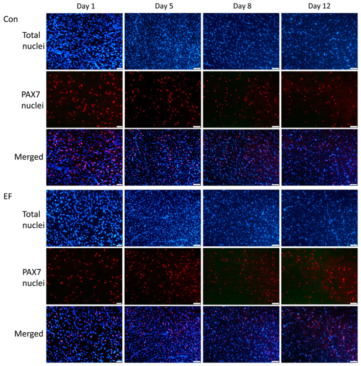
Figure 3.
Immunefluorescence of PAX7 in pectoralis muscle cross sections of untreated control (Con) and E. faecium AL41 (EF) supplemented chicks. Total nuclei were counterstained with DAPI (blue). Merged images indicate nuclear localization of PAX7. Scale bars represent 20 µm at day 1 and 50 µm at other ages.

