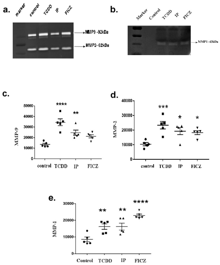
Figure 6.
Activation of AhR induced MMP expression in RAW264.7 cells. In serum-free DMEM media with TCDD (10−8 M), IP (10−7 M), and FICZ (10−7 M) for 24 h, RAW 264.7 cells were incubated. The cell supernatant was collected and subjected to SDS-PAGE (containing 0.1% gelatin or 1% casein) electrophoresis. (a) Representative image of gelatin zymogram. (b) Representative image of casein zymogram. Induction of RAW 264.7 cells with AhR agonists produced (c) MMP-9, (d) MMP-2, (e) MMP-1 expressions. The levels of secreted MMPs are shown as folds of increase compared to the control. Means of five independent experiments +/-SEM were represented in the graph. Unstimulated comparisons to control cultures were depicted by asterisks, such as * p < 0.05, ** p < 0.01, *** p < 0.001 and **** p < 0.0001 by one-way ANOVA analysis.

