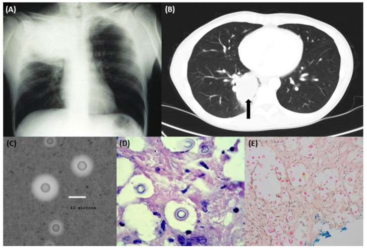
Figure 2.
Representative (A) chest X-ray showing pulmonary Cryptococcus gattii infection with associated Pancoast syndrome (adapted from [33] by courtesy of Oxford Academic); (B) chest high-resolution computed tomography image of a pulmonary cryptococcoma (axial view); (C) India ink stain of Cryptococcus spp.; (D) periodic acid-Schiff and (E) mucicarmine stains of lung tissue with Cryptococcus spp. seen (with capsule staining bright pink in colour).

