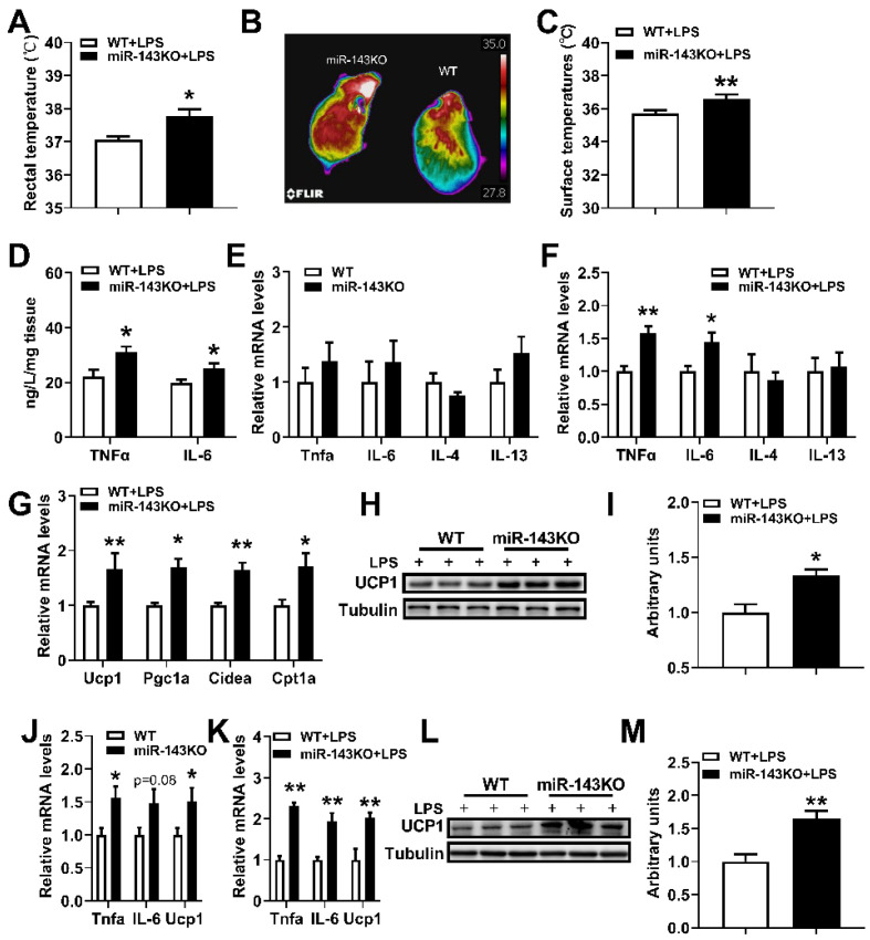
Figure 5.
miR-143KO promotes LPS induced thermogenesis in the BAT. (A) Rectal temperatures of WT and miR-143KO mice treated with 10 μg/kg LPS for 1.5 h. (B,C) Dorsal interscapular surface imaging and temperature. (D) The concentrations of TNFα and IL-6 in the BAT of mice treated with 10 μg/kg LPS for 4 h (n = 6). (E) The inflammation-related mRNA expression levels in the BAT of WT and miR-143KO mice (n = 8). (F,G) The inflammation-related (E) and thermogenic-related (F) mRNA expression levels in the BAT of WT and miR-143KO mice treated with 10 μg/kg LPS for 4 h (n = 6). (H,I) The UCP1 protein levels in the BAT. (J) The mRNA expression levels of TNF-α, IL-6 and UCP1 in the WT and miR-143KO primary brown adipocytes (n = 6). (K–M) The TNFα, IL-6, Ucp1 mRNAs (J), and Ucp1 protein (K,L) expression levels in WT or miR-143KO primary brown adipocytes treated with 0.2 μg/mL LPS. Data are presented as the mean ± SEM. * p < 0.05 vs. controls; ** p < 0.01 vs. controls, as determined by Two-tailed unpaired Student’s t-test.

