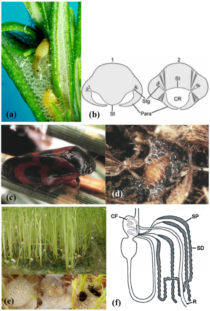
Figure 2.
(a) Philaenus spumarius (Linné, 1758) nymphs inside the foam nest. (b) representation of the abdominal morphology of true spittlebugs nymphs during the production of the foam, in correspondence to the first (1) and fourth (2) urite. St = sternum, Stg = stigmas, Para = paratergal lobes around the respiratory cavity (CR). From Conti et al. [5]. (c) Haematoloma dorsata (Ahrens, 1812) adult and (d) nymph in its foam nest. Images courtesy of José María Cobos, Ministerio de Agricultura, Pesca y Alimentación C/Almagro, Madrid, Spain. (e) Callitettix versicolor (Fabricius, 1794) spittle masses produced by the nymphs at the roots of rice (Oryza sativa L. Poaceae) plants. Images courtesy of Ai-Ping Liang and Xu Chen, Institute of Zoology, Chinese Academy of Sciences, Beijing, China. (f) Representation of the gut complex, including the Malpighian Tubules (MTs), of Homoptera Cercopoidea preimaginal instars. CF = filter chamber, SP = proximal segment of the MTs, SD = distal segment of the MTs, R = rectum. From Conti et al. [5].

