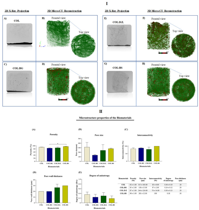
Figure 4.
Micro-CT analysis of the developed COL, COL:BG, COL:D.E., and COL:BS scaffolds, including: (I) Two-dimensional X-ray projections (A,C,E,G) and tridimensional reconstruction images of the frontal and top views (B,D,F,H) for the four the biomaterials; (II) Morphometric properties, namely, (A) Porosity (%), (B) Pore size (µm), (C) Interconnectivity property (%), (D) Pore wall thickness (µm) and (E) Degree of anisotropy. Data are expressed as mean ± SD (n = 3) and the asterisks indicate statistically significant differences (p < 0.05).

