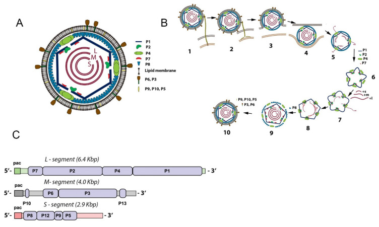
Figure 1.
Schematic organization (A), replication cycle (B), and genome organization (C) of the φ6 virion particle. (A) The inner layer, procapsid (PC), includes the shell composed of 120 copies of the P1 protein or 60 nonsymmetric dimers of P1A/P1B. Inside the shell are proteins P2 and P7 localized at the fivefold axis of symmetry portal and three double-stranded RNA segments (dsRNA) which have low-symmetry quasi-concentric shell organization. The P4 proteins form a hexameric ring around the fivefold axis. The nucleocapsid (NC) includes the PC and a matrix composed of protein P8. The completed virion has an envelope derived from the cellular bilayer lipid membrane and incorporates proteins P3, P6, P9, and P5 randomly distributed. (B) The replication cycle initiates when P3 protein attaches to the host pili followed by pili retraction (1–2). The viral envelope fuses to the host outer membrane (3). The NC enters the periplasmic space and the cytoplasm (4). The P8 matrix disassembles, and transcription begins (5). The PC self-assembles from P1, P2, P4, and P7 proteins (6) and packages the three genome transcripts accompanied by PC expansion (7). ssRNA is replicated to dsRNA (8), and the P8 matrix loosely assembles around the filled PC (9). The cell-derived bilipid membrane is placed around the NC particle mediated by the nonstructural protein P12 (10). This step is followed by cell lysis and virion release. (C) The three ds-RNA genome segments and genes encoding the viral proteins (shown in light-blue color). The packaging pac signal for each strand located at the 5′ end of ds-RNA is visualized as colored rectangles. The replication signal is located at the 3′ end.

