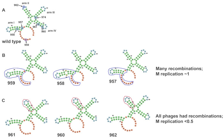
Figure 7.
Alteration of the secondary structure of the 3′ end of the M segment. Introduced deletions of the 3′ end caused changes in the predicted secondary structure. The deleted nucleotides are encircled in blue in (A). (B) The blue outlined arm shows the difference in structure between the wildtype M segment and mutated ones. Changes in the blue outlined arms caused many recombinants in the M segments, but the replication rate of the M segment was nearly the same as of wildtype M. (C) The structure of the red outlined hairpin on these three mutants was different from the second arm in wildtype M. These mutations caused a significant reduction in M replication rate (<5), and all the produced phages were recombinant. Based on from Mindich et al. (1994). The secondary structure was calculated using the forna online RNA secondary structure visualization tool (http://rna.tbi.univie.ac.at/forna/, accessed on 17 August 2022).

