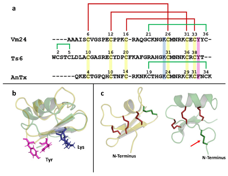
Figure 7.
Structural analysis of Vm24, Ts6, and AnTx. (a) Sequence alignment of Vm24, Ts6, and AnTx. Conserved Cys between the three peptides are highlighted in yellow; the functional dyad is highlighted in blue and pink. Conserved disulfide bridges are shown in red; the extra disulfide bridges are shown in green. (b) Structural alignment between Vm24 and Ts6. Vm24 structure is shown in yellow (PDB:2K9O); Ts6 structure is shown in green (PDB:1C56); functional dyads are shown in the structural alignment as sticks, lysine (blue) and tyrosine (pink). (c) Disulfide pattern of Vm24 and Ts6. Conserved disulfide bridges are shown in red sticks; the other disulfide bridges are shown in green. The red arrow points to the disulfide bridge found in the N-terminus of Ts6.

