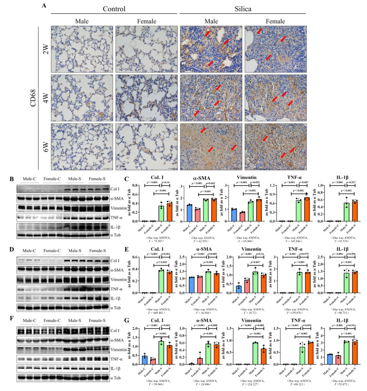
Figure 4.
Western blot of CD68, COL I, vimentin, and α-SMA in male and female mice exposed to silica for two, four, six weeks, and the control groups. (A) Immunohistochemical staining was used to detect the expression of CD68 in the mouse lung tissues, arrow: CD68-positive cells (brown) (scale bar = 50 µm). (B–G) Expression levels of Col I, α-SMA, vimentin, IL 6 and IL 1β in the lungs of mice were measured using the western blot. Data are presented as the mean ± SD, n = 5 per group ((B,C)—2W, (D,E)—4W, (F,G)—6W).

