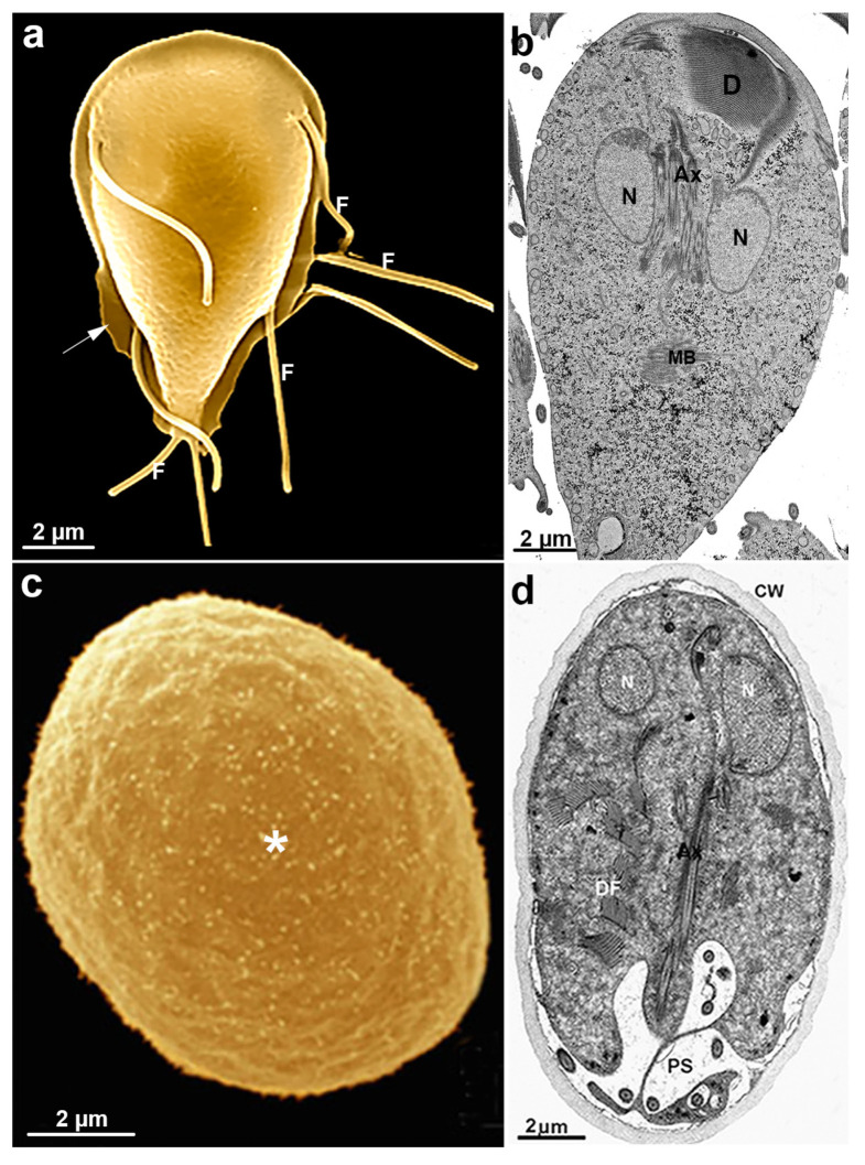
Figure 11.
G. intestinalis trophozoite and cyst: (a) scanning electron microscopy of the parasite. The four flagella (F) pairs, ventrolateral flange (arrow), and dorsal surface are observed; (b) transmission electron microscopy of a trophozoite showing internal structures as the nuclei (N), median body (MB), ventral disc (D), and axonemes (Ax); (c) scanning electron microscopy image shows an oval appearance of the cyst and a fibrillar material (*) deposited over the cell; and (d) transmission electron microscopy of the cyst. The nuclei (N), disc fragments (DF), and axonemes (Ax) are observed, as well as the cyst wall (CW) and peritrophic space (PS). (a,b) (Gadelha and Benchimol, unpublished); (c,d) (After Ref. [78]).

