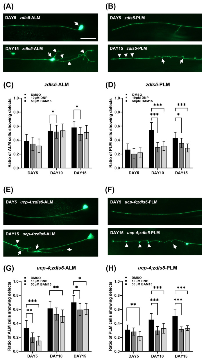
Figure 1.
BAM15 attenuates neuronal defects in aged C. elegans. (A,B) Representative images of mechanosensory neurons in zdIs5 [Pmec-4::GFP] animals on Day 1 and Day 15; ALM and PLM, respectively. Arrows indicate soma and arrowheads indicate soma outgrowth and branching in ALM (A). Arrows indicate blebs and arrowheads indicate wavy processing in PLM (B). Scale bar = 50 µm. (C,D) ALM and PLM neuronal defects are presented as a ratio of the total ALM- and PLM-scored neurons in animals treated with 10 µM DNP and 50 µM BAM15 on Days 5, 10, and 15. (E,F) Representative images of mechanosensory neurons in ucp-4;zdIs5 animals on Days 1 and 15; ALM and PLM, respectively. Arrows indicate neuronal sprouting and arrowhead indicates a bleb in ALM (E). An arrow indicates branching and arrowheads indicate blebs (F). (G,H) ALM and PLM neuronal defects are presented as a ratio of the total ALM- and PLM-scored neurons in ucp-4;zdIs5 animals treated with 10 µM DNP and 50 µM BAM15 on Days 5, 10, and 15. ALM, anterior lateral microtubules; PLM, posterior later microtubules. Data are represented as means of three independent experiments. Error bars represent the SEM. n = 80~120 worms per treatment in each experiment. * p < 0.05, ** p < 0.01, *** p < 0.001. Unpaired Student’s t-tests were performed to compare the DMSO control and treatments.

