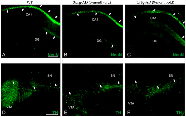
Figure 2.
Biomarker expression in transgenic mice with AD-like neuropathology. Photomicrographs of brain sections from WT (n = 3–4) and APP/BIN1/COPS5 3xTg-AD (n = 3–4) mice showing histopathological lesions identified with immunohistochemical biomarkers. (A–C) Transverse sections of the dentate gyrus (DG) and CA1 area of the hippocampus in WT- (A) and APP/BIN1/COPS5 3xTg-AD mice (B,C), showing strong differences in NeuN immunoreactivity (white arrows). (D–F) VTA highlighting the notable reduction in TH-stained cells (white arrows) in the female APP/BIN1/COPS5 3xTg-AD mouse brain (E,F), compared to WT mice (D). Area fraction analyses of the CA1 subfield (G) and DG (H) of the hippocampus, and VTA in the midbrain (I), found reductions in NeuN and TH immunoreactivities, respectively, in 3-m.o.- and 9-m.o.-3xTg-AD mice versus WT animals. Data represent mean ± S.E.M. One-way ANOVA followed by Tukey’s post hoc comparisons, * p < 0.05, ** p < 0.01, *** p < 0.001. Scale bars, 100 μm. 3xTg-AD, triple-transgenic mouse model of AD; CA1, cornu Ammonis 1; DG, dentate gyrus; m.o., month-old; NeuN, neuronal nuclear protein; SN, substantia nigra; TH, tyrosine hydroxylase; VTA, ventral tegmental area; WT, wild-type.


