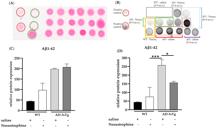
Figure 3.
Aβ1-42 levels in young (3–4-month-old) and older (8–9-month-old) wild-type- and AD transgenic mice. Hippocampal protein (1 µg/µL) from wild-type saline-treated young (n = 3, black box in (B)) and older mice (n = 3, black box in (B)), wild-type Nosustrophine-treated young (n = 2, orange box in (B)) and older mice (n = 3, blue box in (B)), APP/BIN1/COPS5 3xTg saline-treated young (n = 2, purple square bracket in (B)) and older mice (n = 4, red boxes in (B)), and APP/BIN1/COPS5 3xTg Nosustrophine-treated young (n = 2, grey box in (B)) and older mice (n = 2, green box in (B)), Aβ-peptide (1-42) (0.5- and 1 µg/µL) and BSA (0.01–1 µg/µL) were spotted on a nitrocellulose membrane and stained with Ponceau S (A). The membrane was then incubated with an antibody (clone 12F4) against mouse Aβ1-42 (B). (C) Quantification of Aβ1-42 levels in young (3–4-month-old) and (D) older (8–9-month-old) mice. Data represent mean ± S.E.M. One-way ANOVA followed by Tukey’s post hoc comparisons, * p < 0.05, *** p < 0.001. AD-3xTg, triple-transgenic mouse model of AD; Aβ, amyloid-beta; AD, Alzheimer’s disease; BSA, bovine serum albumin; m.o., month-old; No-us., Nosustrophine; WT, wild-type. Red circles in A, positive controls (Aβ-peptide 1-42); green circles in A, negative controls (BSA).

