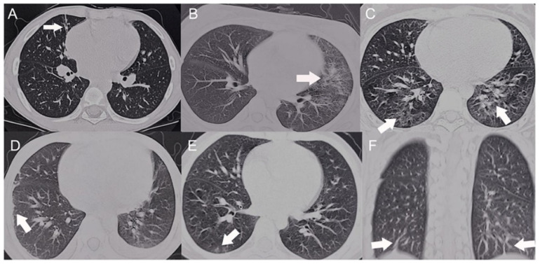
Figure 2.
HRCT scans of the SLE patients with pulmonary involvement. Arrow indicate the shape of lung lesion of HRCT (A) Fibrotic streak. (B) Reticular pattern. (C) Mosaic perfusion. (D) Pleural thickening. (E) Ground glass opacity. (F) Subpleural interlobular septal thickening (The CC-BY Creative Commons attribution license, Dai G, Li L, Wang T, Jiang W, Ma J, Yan Y, Chen Z. Pulmonary Involvement in Children With Systemic Lupus Erythematosus. Front Pediatr. 2021 Feb 2; 8:617137. doi: 10.3389/fped.2020.617137. PMID: 33604317; PMCID: PMC7884320) [69].

