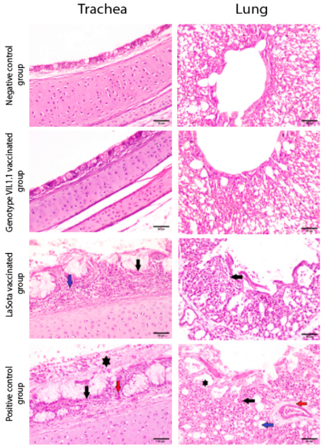
Figure 7.

Photomicrographs representing H&E stained sections of tracheas and lungs collected from genotype VII.1.1 and LaSota vaccinated- challenged chicks with virulent NDV compared to mock chicks (negative control) and positive control groups (non-vaccinated challenged chicks). Non-vaccinated- non-challenged chicks and genotype VII.1.1 vaccinated- challenged chicks showing normal histological architecture for Tracheas and normal histological architecture of parabronchus and air capillaries in lungs. On the other hand, LaSota vaccinated challenged chicks showed necrosis of lamina epithelialis and mucosal glands (black arrow) associated with mononuclear cells infiltration in lamina propria (blue arrow) in Trachea and inflammatory cells infiltration (black arrow) in lungs. In addition, non-vaccinated-challenged chicks (positive control group) showing multifocal necrosis of lamina epithelialis (black arrow), congestion (red arrow) and accumulation of mucous exudate in the tracheal lumen (asterisk) in Trachea and showing inflammatory cells infiltration in the air capillaries (black arrow), perivascular edema (red arrow), dilatation of atria (asterisk) and dilatation of air capillaries (blue arrow) in lungs (scale bar 50 µm).
