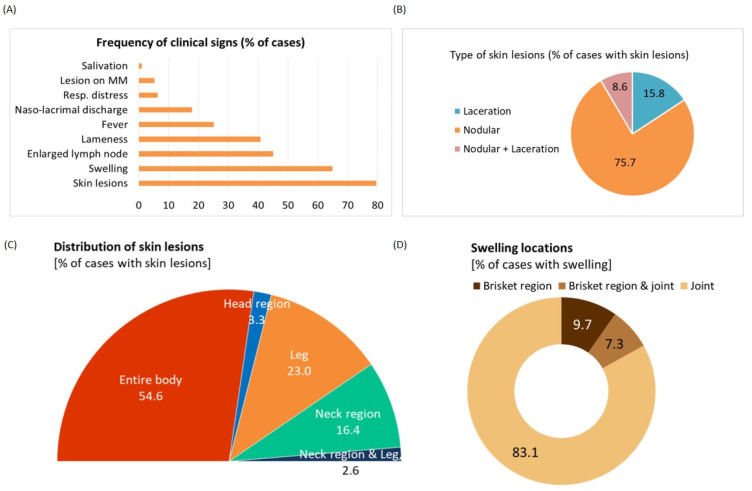
Figure 1.
Clinical observations and distribution of lesions of the affected cattle population. (A) Frequency of clinical signs shown by the affected cattle. (B) Pie chart showing the type of skin lesions that were classified as nodular, laceration, and both. (C) Half pie chart showing the distribution of skin lesions in different parts of the body. (D) Pie chart revealing the occurrence of swelling in different body parts.

