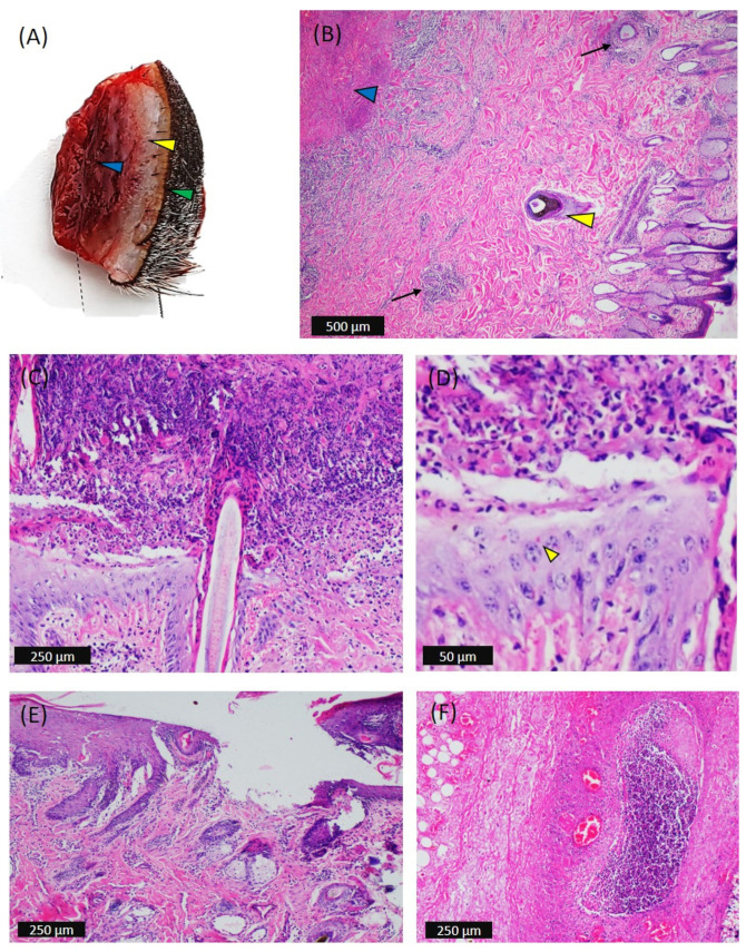
Figure 3.
Gross and microscopic pathology of the skin nodules in affected cattle. (A) Biopsy of a skin nodule that was 2 cm in diameter that provided gritty sound when cut and showed hemorrhagic subcutis (blue arrowhead), pale dermis (yellow arrowhead), and haired epidermis (green arrowhead). (B) Epidermis: epidermal cytoplasmic swelling and mononuclear infiltrate. Dermis: diffuse proliferation of mononuclear cells between the dense irregular connective tissue and reticular layers and in the perivascular space (black arrow). Sweat and sebaceous glands were found dilated, and there was intracellular edema of sebaceous gland cells. The hair follicle matrix epithelia were found to be highly hyperplastic (yellow arrowhead). Subcutis: presence of an infarct (blue arrowhead). (C) Epidermis and dermis: vacuolation, swelling, and adhesion of scabs with proliferative stratum basale, intraepidermal necrosis, and accumulation of scale crust leaving ulcer underneath; in addition, the dermis was hemorrhagic, edematous, and infiltrated with mononuclear cells. (D) Occasionally, keratinocytes were consistent with intracytoplasmic inclusion bodies (yellow arrowhead). (E) Vacuolation and swelling of keratinocytes, epidermal proliferation of basal cells, infiltration of round histiocytic cells in the reticular layer of skin, swelling, and vacuolation of glandular epithelium and focal ulceration. (F) Subcutis: presence of focal aggression of mononuclear cells, congestion, necrosis, and lysis of subcutaneous fat cells.

