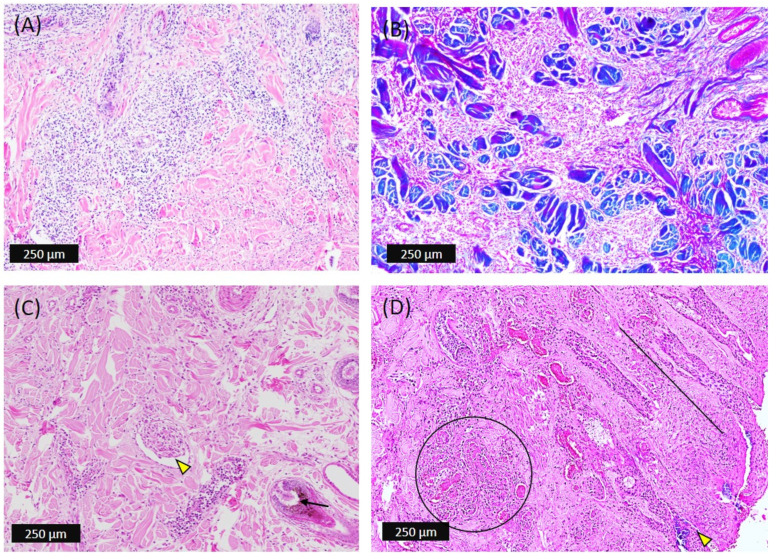
Figure 4.
Microscopic pathology of the skin nodules developed in dermis of affected cattle. (A) Diffuse to nodular proliferation of mononuclear cells in the dense irregular connective tissue layer of dermis as well as centering the blood vessels. (B) Abundance of connective tissue fibers (blue) after using Masson’s trichrome stain. (C) The inflammatory cells adhered to lumen and around blood vessels (yellow arrowhead); the hair follicles’ epithelial cells are degenerated and hyperplastic (black arrow). (D) Dermis showing massive necrotizing dermatitis with multiple infarcts (black circle) and formation of excretory sinuses (black line) with invaded bacteria (yellow arrowhead).

