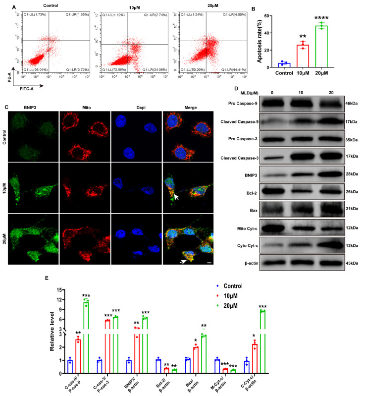
Figure 3.
MLD induced the apoptosis of HepG2 cells by promoting the expression of BNIP3. (A) MLD induced apoptosis, as detected by Annexin V-FITC/PI double staining assay. (B) Quantitative analysis of the apoptosis. (C) The protein level of BNIP3 was detected by immunofluorescence. Scale bars: 50 μm. Arrow: the colocalization of BNIP3 and mitochondria. (D) Levels of the pro-caspase-9, cleaved caspase-9, pro-caspase-3, cleaved caspase-3, BNIP3, Bcl-2, Bax, Mito Cyt-c, and Cyto Cyt-c proteins in the different groups were analyzed by Western blotting. (E) The quantitative analysis of relative protein levels. The results are representative of three independent experiments and are expressed as the mean ± SD. * p < 0.05, ** p < 0.01, *** p < 0.001, and **** p < 0.0001 compared with the control group.

