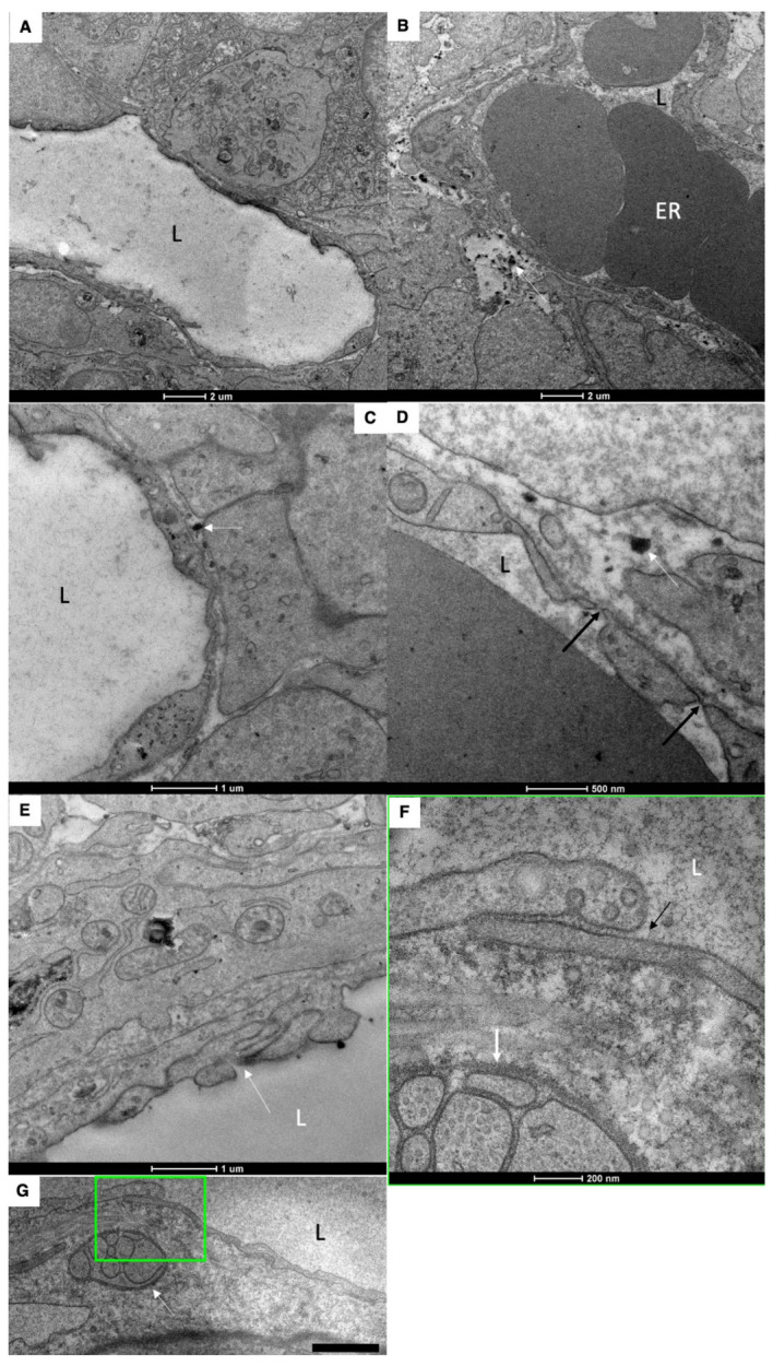
Figure 4.

Structure of blood and lymph capillaries in newborn rats before their first feeding. (A–C) Blood capillary. Low number of fenestrae in capillary wall. (D) Low density of fenestrae (black arrows). White arrows in (B–D) show membrane remnants in the interstitial space. (E–G) EM images of the LC wall. White arrow in (E) shows complex inter-endothelial contact. White arrows in (F,G) indicate nervous terminal. The square box with green border it enlarged in (F). (F) Enlarged area inside the square box with green borders in (G). Black arrows indicate the simple tile-like contact between endothelial cells of LC. Low number of caveolae. L, lumen of blood and lymph capillaries. ER, erythrocytes. Scale bars: 990 nm (G). In (A–F), scale bars are indicated below images.
