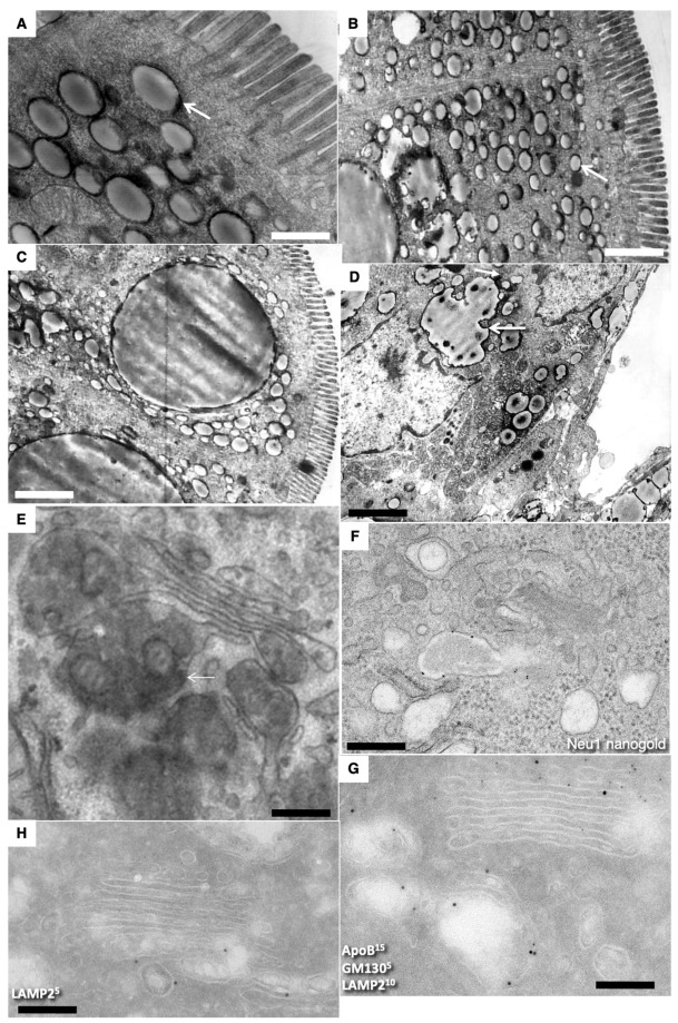
Figure 5.
Structure of enterocytes in newborn rats after their first feeding. (A–C) Formation of large lipid droplets (white arrows) in the cytoplasm. (D) Formation of “lipid lakes” (black arrows) between enterocytes after the first feeding of a newborn rat. (E) Overloading of the GC. (F) Large ChMs in post-Golgi compartment positive for Nau1. (G,H) Labeling for LAMP2, GM130 and ApoB of post-Golgi carriers containing large ChMs. Scale bars: 510 nm (A); 1020 nm (B); 1.3 µm (C); 1025 nm (D); 240 nm (E); 340 nm (F).

