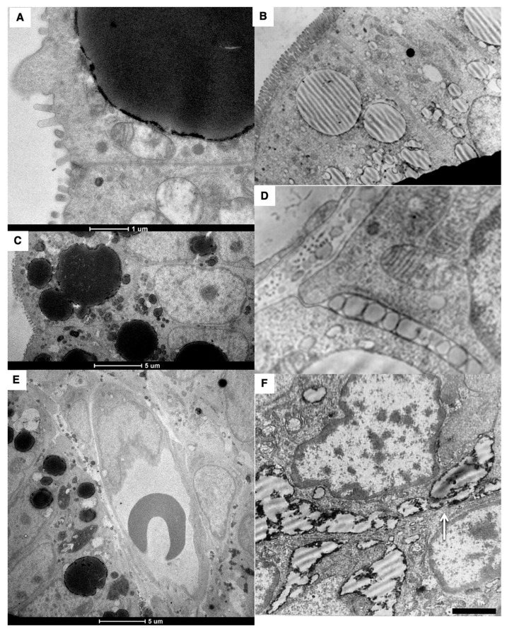
Figure 6.
Structure of small intestine enterocytes in newborn rats after their first feeding. (A–C) Formation of huge droplets and “lipid lakes” in enterocytes of newborn rats after the first feeding. (A,C) Thick (200 nm) sections. (D) Large ChMs between enterocytes. (E) The lumen of the blood capillary with the erythrocytes inside its lumen does not contain chylomicrons. (F) “Lipid lakes” between enterocytes. Scale bars: 1050 nm (B); 290 nm (D); 610 nm (F). In (A,C,E), scale bars are indicated below each image.

