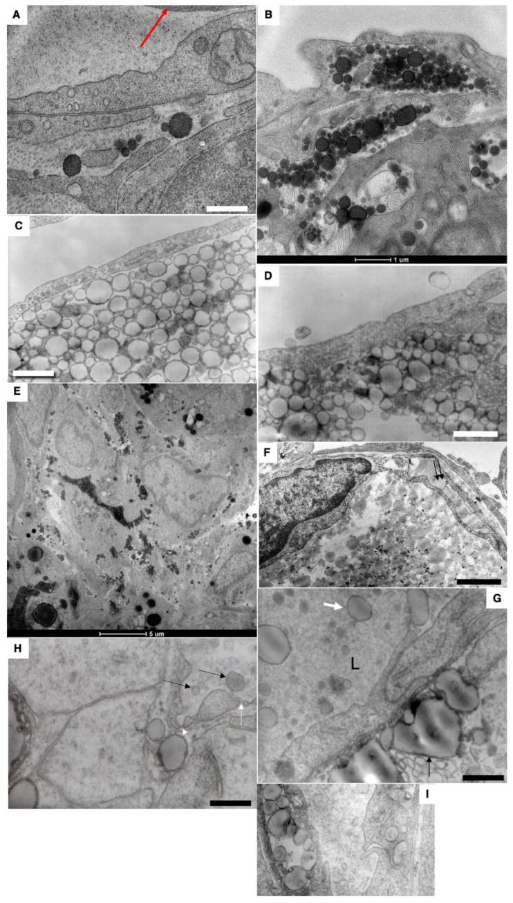
Figure 7.

Passage (presumably) of large chylomicrons through intercellular spaces. (A) Large chylomicrons are not captured by blood capillaries (the red arrow shows erythrocyte). (B–D) Accumulation of chylomicrons in the interstitial space. (E). Accumulation of chylomicrons between enterocytes. (F,G) Uptake of chylomicrons into the lymphatic capillary. Double arrows in (F) show large chylomicron in the inter-endothelial contact. (G) Large ChMs in the lumen of the lymphatic capillary. (H) Rarely, ChMs (shown by black arrows) were seen in the blood capillary, in the wall of which single fenestrae are visible (shown by white arrows). (I) Chylomicrons (to the right) inside the vacuoles within cytoplasm of endothelial cells of lymphatic capillary. Scale bars: 595 nm (A,H); 610 nm (C): 460 nm (D,I); 900 nm (F); 570 nm (G). In (B,E), scale bars are indicated below images.
