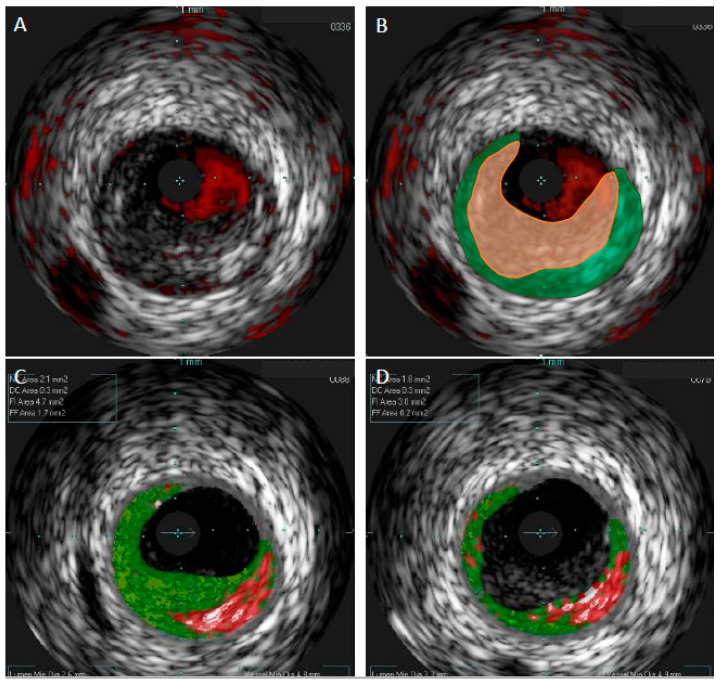
Figure 2.
Comparison of gray-scale intravascular ultrasound and virtual histology intravascular ultrasound in patient with acute coronary syndrome. In gray-scale intravascular ultrasound (GS-IVUS), cross-section with lumen narrowing could be interpreted as soft plaque (A). However, on live image, motion and oscillation of the “plaque” were visible—image typical for thrombus. After postprocessing, thrombus (orange zone) was separated from true plaque (green zone) in GS-IVUS (B). In the same patient virtual histology intravascular ultrasound (VH-IVUS) marked thrombus as fibrotic plaque (C). Only after postprocessing could visualization of real borders of the plaque be presented (D).

