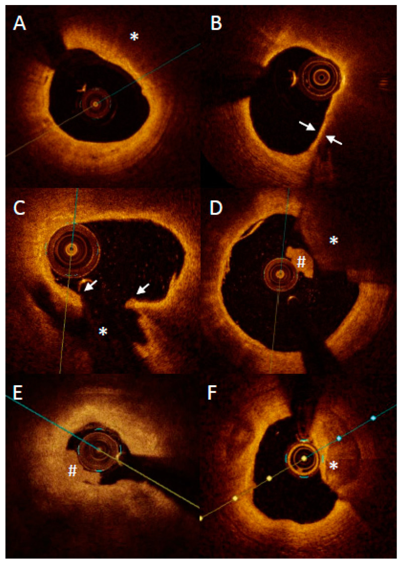
Figure 4.
Representative images of optical coherence tomography findings in patients with acute myocardial infarction. Lipid plaque is characterized as signa poor regions (asterisk) with overlying signal-rich bands (A). Thin-cap fibroatheroma is defined as a lipid plaque occupying more than >90° in circumference and with fibrous cap thickness (arrows) less than a set threshold (usually 65 μm or 80 μm) (B). Plaque rupture is defined as disruption of fibrous cap (arrows) with visible cavity within the plaque ((C) asterisk). Red thrombus is described as highly backscattering structure with high attenuation ((D) asterisk), whereas white thrombus is less backscattering and has lower attenuation ((D,E) #). Erosion is described as presence of attached thrombus (usually white; #) overlying an intact and visualized plaque (E). Calcification protruding to the lumen is described as calcific nodule ((F) asterisk).

