Figure 1.

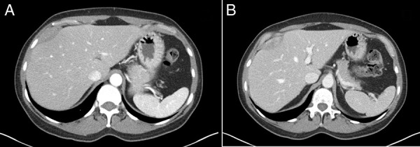
Contrast-enhanced 3-phase computed tomography demonstrating a 1.9 × 5.0 × 6.3 cm fusiform mass along the capsule of the liver anterior to hepatic segments 8/4a with mild arterial enhancement (A) and progressive delayed enhancement (B).

Contrast-enhanced 3-phase computed tomography demonstrating a 1.9 × 5.0 × 6.3 cm fusiform mass along the capsule of the liver anterior to hepatic segments 8/4a with mild arterial enhancement (A) and progressive delayed enhancement (B).