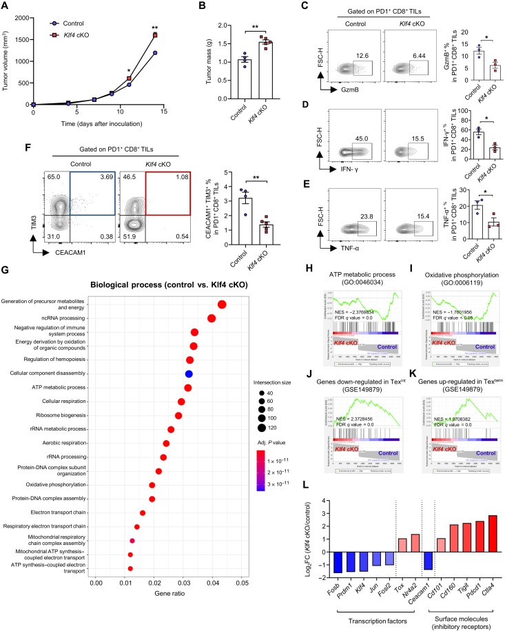
Fig. 5. KLF4 deficiency impairs CD8 T cell differentiation into transitory effector subsets and the antitumor function.
(A to L) MC38 cells were subcutaneously injected into the right flank of control/Klf4 cKO mice. CD8+ TILs were analyzed on day 14. (A) Tumor growth curve of control (n = 4)/Klf4 cKO (n = 5) mice. (B) Histogram of tumor mass of control (n = 4)/Klf4 cKO (n = 5) mice. (C to E) Representative flow cytometry plot and the proportion of (C) GzmB+, (D) IFN-γ+, and (E) TNF-α+ cells in PD1+CD8+ TILs from control/Klf4 cKO mice (n = 3 per group). (F) Representative flow cytometry plot and the proportion of CEACAM1+TIM3+ cells in PD1+CD8+ TILs from control (n = 4)/Klf4 cKO (n = 5) mice. (G to L) PD1+CD8+ TILs from tumor tissues of four control mice and six Klf4 cKO mice were pooled on day 14, and SMART-seq was performed. (G) GO (GO biological process) analysis of DEGs. rRNA, ribosomal RNA. (H to K) GSEA between control and Klf4 cKO PD1+CD8+ TILs using gene sets of (H) adenosine triphosphate (ATP) metabolic process, (I) oxidative phosphorylation, (J) genes down-regulated in Texint, and (K) genes up-regulated in Texterm. (L) Histogram of fold change of genes between control and Klf4 cKO PD1+CD8+ TILs. (A to F) Data are means ± SEM. Statistical analysis was performed using Student’s t test. *P < 0.05; **P < 0.01.

