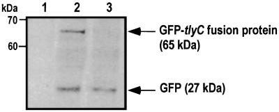
FIG. 4.
Detection of a GFP-tlyC fusion protein by immunoblotting with the anti-GFP monoclonal antibody. Lanes 1 to 3 contain nonhemolytic P. mirabilis WPM111/hpmA, P. mirabilis WPM111/hpmA complemented with GFP-tlyC, and P. mirabilis WPM111/hpmA transformed with GFPuv only, respectively. GFP-tlyC fusion protein was detected at 65 kDa.

