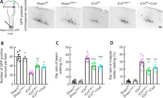
Figure 8.

CypD deficiency and CsA treatment protect the CST and alleviate motor dysfunction after ICH.
(A) Diagram of retrograde tracking of the CST (left) and GFP-positive corticospinal neurons (right, Bregma: –0.7 mm) in each group. The number of retrograde labeled GFP-positive corticospinal neurons in the ICHCypD–/– and ICHWT + CsA groups was greater than that in the ICHWT group. Scale bar: 100 μm. (B) Number of labeled corticospinal neurons in each group. (C, D) The slip ratio of the contralateral limbs in beam walking (C) and ladder rung walking (D) at 3 days post-ICH in each group. Data are shown as the mean ± SEM (n = 6–8 animals for each group). *P < 0.05, **P < 0.01, ***P < 0.001, vs. ICHWT group (one-way analysis of variance followed by Tukey’s post hoc test). CsA: Cyclosporin A; CST: corticospinal tract; CypD: cyclophilin D; GFP: green fluorescent protein; ICH: intracerebral hemorrhage; WT: wild type.
