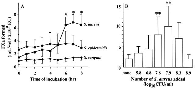
FIG. 3.
(A) Course of TFA in bacteria-infected ECs. Monolayers of 2 × 10⁵ ECs were incubated at 37°C with 3 × 10⁷ to 5 × 10⁷ S. aureus, S. sanguis, or S. epidermidis organisms. At the indicated time points, the monolayers were washed and assessed for TFA by measuring FVIIa-dependent FX activation as described in Materials and Methods. ∗, P < 0.001 versus 0 h. (B) Dose dependency of the S. aureus-induced endothelial TFA. Monolayers of ∼2 × 10⁵ ECs were incubated for 7 h at 37°C with medium alone (none) or 1 ml of the indicated S. aureus numbers. After washing, TFA was assessed as described above. ∗∗, P < 0.05 versus none. Values represent the means ± standard deviations of five (A) or four (B) experiments with ECs from different donors.

