Figure 3.
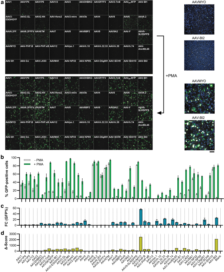
Transduction efficiency on THP-1 cells dependent on PMA stimulation. Sixty thousand THP-1 cells were seeded into 96-well plates and conditionally stimulated with 20 ng/mL of PMA, followed by cultivation in regular media. Twenty-four hours after seeding, cells were transduced with the AAV panel (2 × 10⁹ vg per AAV variant). Three days later, images were taken by (a) high-content fluorescence microscopy and (b) GFP-positive cells were quantified by semiautomated image analysis. The fold change in the number of GFP-expressing cells between PMA-free and PMA-stimulated conditions is depicted in (c) and a composite Δ-score (fold change × %GFP-positive cells) is shown in (d). Enlarged images of the cells transduced with AAVMYO and AAV-BI2 under PMA-free (top) and PMA-treated (bottom) conditions are shown in the right part of (a). Blue staining indicates Hoechst33342-stained nuclei. Scale bar = 100 μm. n = 3 replicate plates each, mean ± SD. PMA, phorbol 12-myristate 13-acetate.

