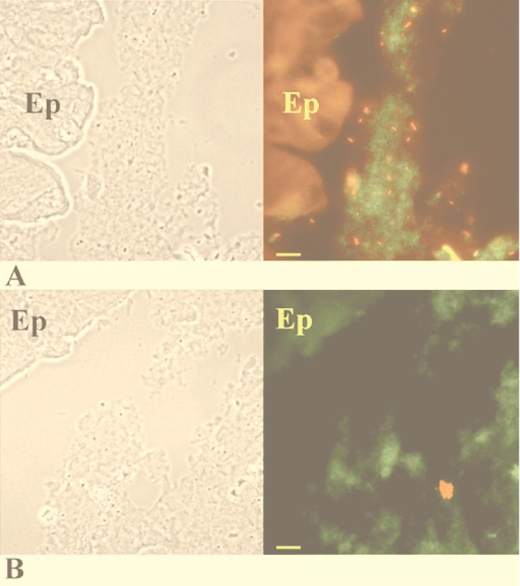
FIG. 4.
Colonic sections of mice fed wild-type K. pneumoniae LM21 (A) and the K. pneumoniae LM21(cps) capsule-defective mutant (B) on day 20 after infection. (Left) Phase-contrast picture of the corresponding area of in situ hybridization. (Right) In situ hybridization with fluorescence-labeled oligonucleotide probes. K. pneumoniae bacteria appear red, while other eubacteria appear green. The colonic epithelium (Ep) is fluorescent because of the autofluorescence of the eucaryotic tissue. Bars, 10 μm.

