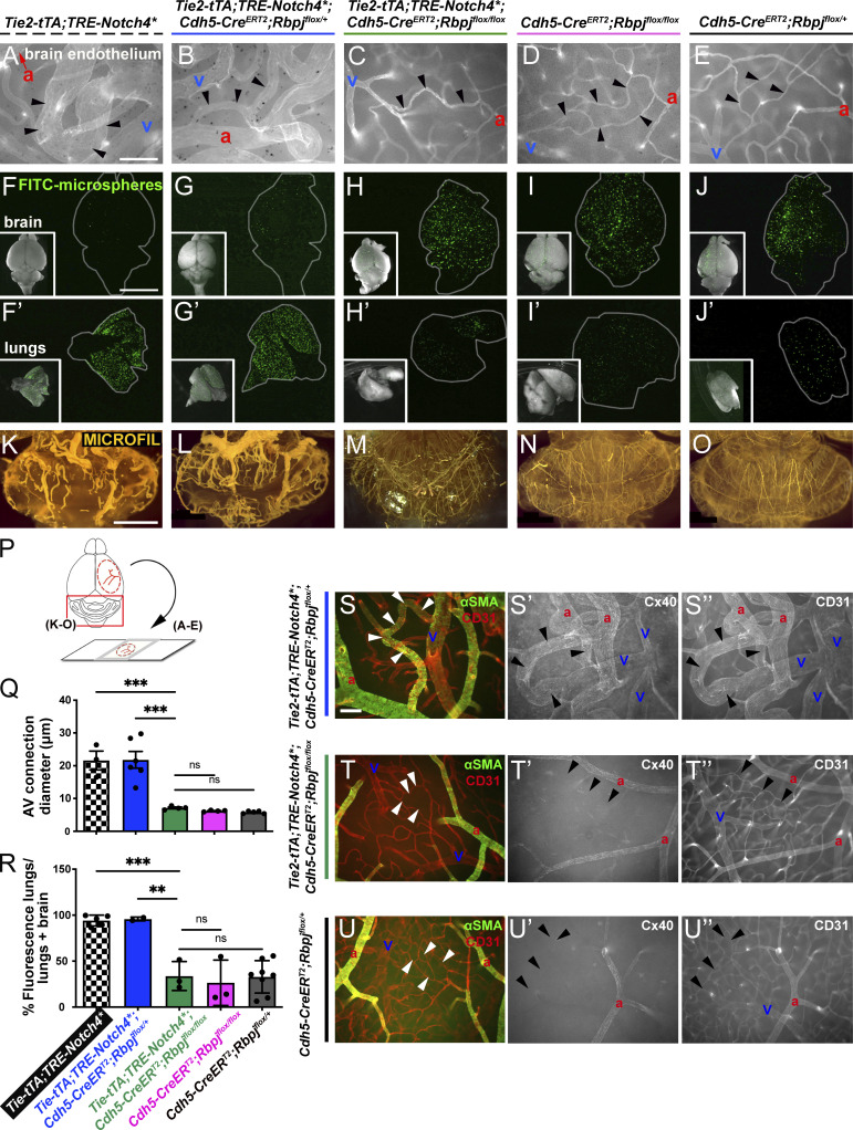
Figure 3.
Endothelial deletion of Rbpj from P16 normalized brain AVM phenotypes and restored arterial identity, in AV connections and veins, in Notch4*tetEC mice. (A–E) Whole-mount frontal cortex with FITC-lectin+ ECs to highlight vessels. We found diameters of AV connections were significantly reduced in Notch4*tetEC;RbpjiΔEC brains (7.14 ± 0.53 μm; N = 4 mice, 185 connections), as compared with Notch4*tetEC (21.61 ± 4.97 μm; N = 3 mice, 116 connections) or Notch4*tetEC;RbpjiΔEC-het (21.83 ± 6.24 μm; N = 6 mice, 315 connections) brains. Importantly, AV connection diameters in Notch4*tetEC;RbpjiΔEC mice were not significantly different from either RbpjiΔEC mice (6.22 ± 0.23 μm; N = 4 mice, 202 connections) or negative controls (5.79 ± 0.38 μm; N = 5 mice, 259 connections). Arrowheads indicate AV connections. a, artery; v, vein. AV connection diameters were quantified in (Q). Mice from eight litters or eight independently repeated experiments. (F–J′) Microspheres passed through AV shunts in Notch4*tetEC (94.18 ± 5.86%, N = 5) and Notch4*tetEC;RbpjiΔEC-het (95.83 ± 2.06%, N = 3) brains but lodged in Notch4*tetEC;RbpjiΔEC (33.81 ± 28.65%, N = 3) brain AV connections, as well as in AV connections from RbpjiΔEC (26.54 ± 24.67%, N = 3) and negative control (32.91 ± 17.66%, N = 8) brains. Microsphere passage quantified in (R). Mice from seven litters. (K–O) MICROFIL casting of cerebellar vasculature. Enlarged and tortuous vessels were cast in Notch4*tetEC (N = 4) and Notch4*tetEC;RbpjiΔEC-het (N = 5) brains but were not readily observed in Notch4*tetEC;RbpjiΔEC (N = 4), RbpjiΔEC (N = 7), or negative control (N = 6) brains. 10 independently repeated experiments. (P) Schematic indicates whole brain regions shown in panels. Scale bars: 100 μm in A–E; 5 mm in F–J′; 2 mm in K–O. (S–U″) Whole-mount frontal cortex was immunostained against CD31 (to label ECs) and αSMA (S–U) or Cx40 (S′–U′) to label arterial ECs or CD31 (S″–U″) to label all ECs. White or black arrowheads indicate AV connections. a, artery; v, vein. In Notch4*tetEC;RbpjiΔEC-het cortex, αSMA (S) and Cx40 (S′) expression extends beyond arteries, throughout AV shunts (arrowheads) and into veins. In both Notch4*tetEC;RbpjiΔEC and negative control cortex, arterial markers αSMA (T–U) and Cx40 (T′–U′) are expressed in arteries but not in AV connections (arrowheads) or veins. N = 3 mice for each genotype. Three independently repeated experiments. Scale bars: 100 μm. **P<0.01; ***P<0.001.

