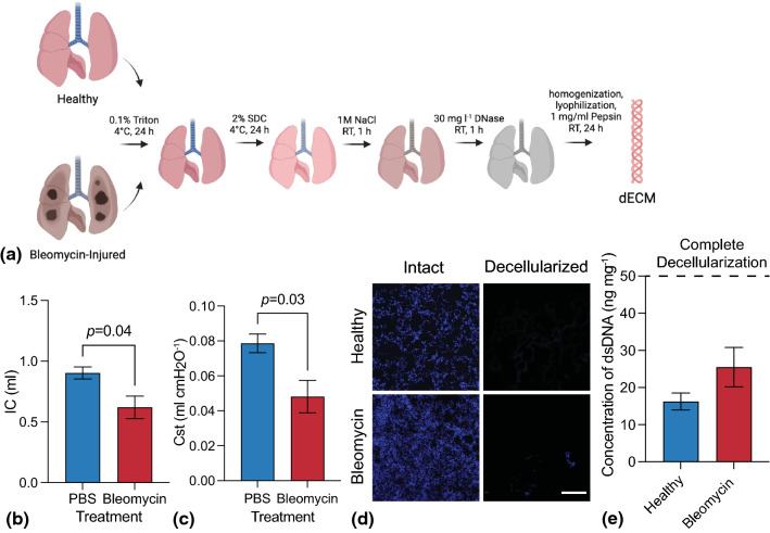
Figure 1.
(a) Schematic representing the lung decellularization process for both healthy and bleomycin-injured lungs. Lungs were sequentially perfused with detergents, salts, and enzymes before being enzymatically digested and lyophilized. (b) A significant decrease in (b) inspiratory capacity and (c) quasi-static compliance was measured in bleomycin-injured lungs versus controls, indicating reduced air volume in lung due to fibrotic tissue accumulation (N = 5–8; Student’s t-test). (d) Representative fluorescent microscopy images of cell nuclei (blue; DAPI) before and after decellularization showed nearly complete removal of cells in healthy and bleomycin-injured mouse lung samples (N = 3, Scale bar 200 µm). (e) Quantification of dsDNA concentration demonstrated decellularized scaffolds containing average dsDNA amounts that were significantly below the threshold for complete decellularization (N = 3).

