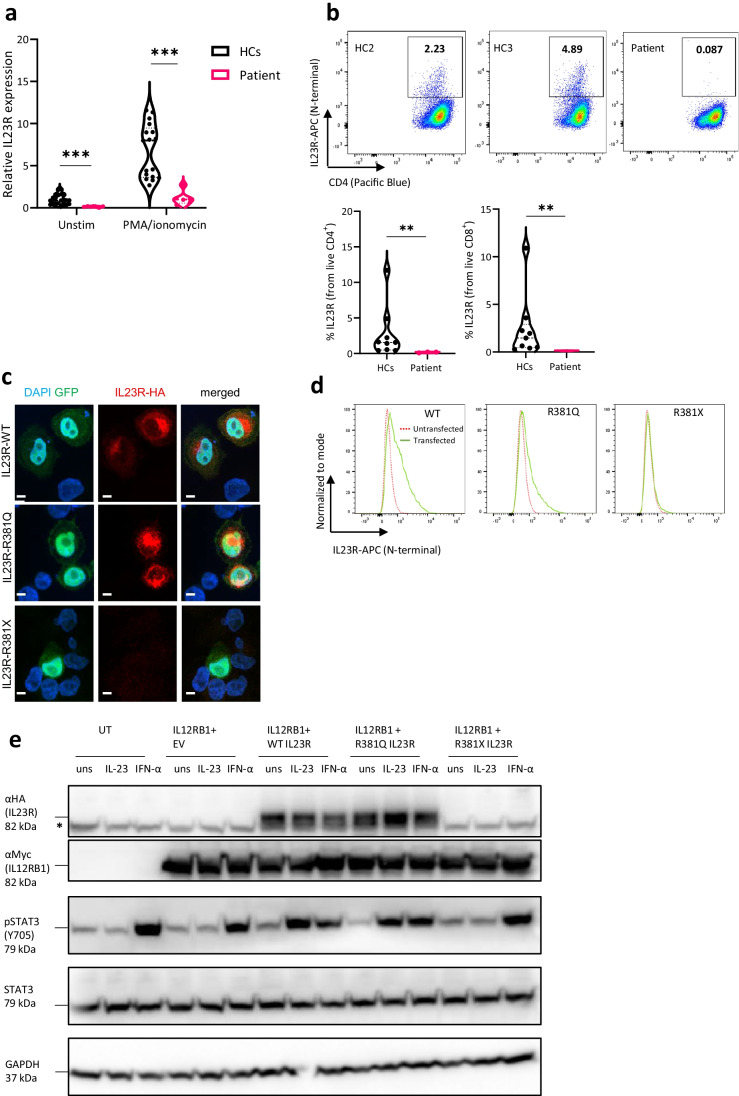
Fig. 2.
Near absence of IL23R expression in patient PBMCs and absent activity of overexpressed IL23R R381X in HeLa cells. a qPCR for IL23R corrected for GAPDH and normalized to HCs on PBMCs. PMA/ionomycin = phorbol 12-myristate 13-acetate/ionomycin (4 h, 25 ng/mL + 1 µg/mL). Data pooled from 6 independent experiments, comparison by unpaired Student T-test. b IL23R surface (N-terminal antibody) staining on CD4+ T cells from HCs (n = 9, 2 shown) and patient (n = 1, 3 biological replicates, 1 shown). Fluorescence minus one (FMO) IL23R was used for proper gating. Percentage of IL23R+ cells from CD4+ and CD8.+ live cells, data pooled from 2 independent experiments. Mann–Whitney U test was used for comparison. c Confocal images of transfected (GFP) HeLa cells with WT, R381Q or R381X IL23R (C-terminally 3xHA tagged IL23R IRES GFP constructs), representative of 2 independent experiments. Counterstaining with DAPI. Scale bar is 5 μm. d IL23R surface (N-terminal antibody) staining on transfected HeLa cells with WT, R381Q or R381X IL23R (C-terminally 3xHA tagged IL23R IRES GFP constructs), representative of 2 independent experiments. e STAT3 phosphorylation in HeLa cells untransfected or cotransfected with IL12RB1, empty vector GFP (EV), IL23R WT, IL23R R381Q or IL23R R381X plasmids, unstimulated or stimulated with IL23 (100 ng/mL) or IFN-α (10 ng/mL) for 1 h, representative blot of 3 independent experiments. * indicates the STAT-3 band under αHA

