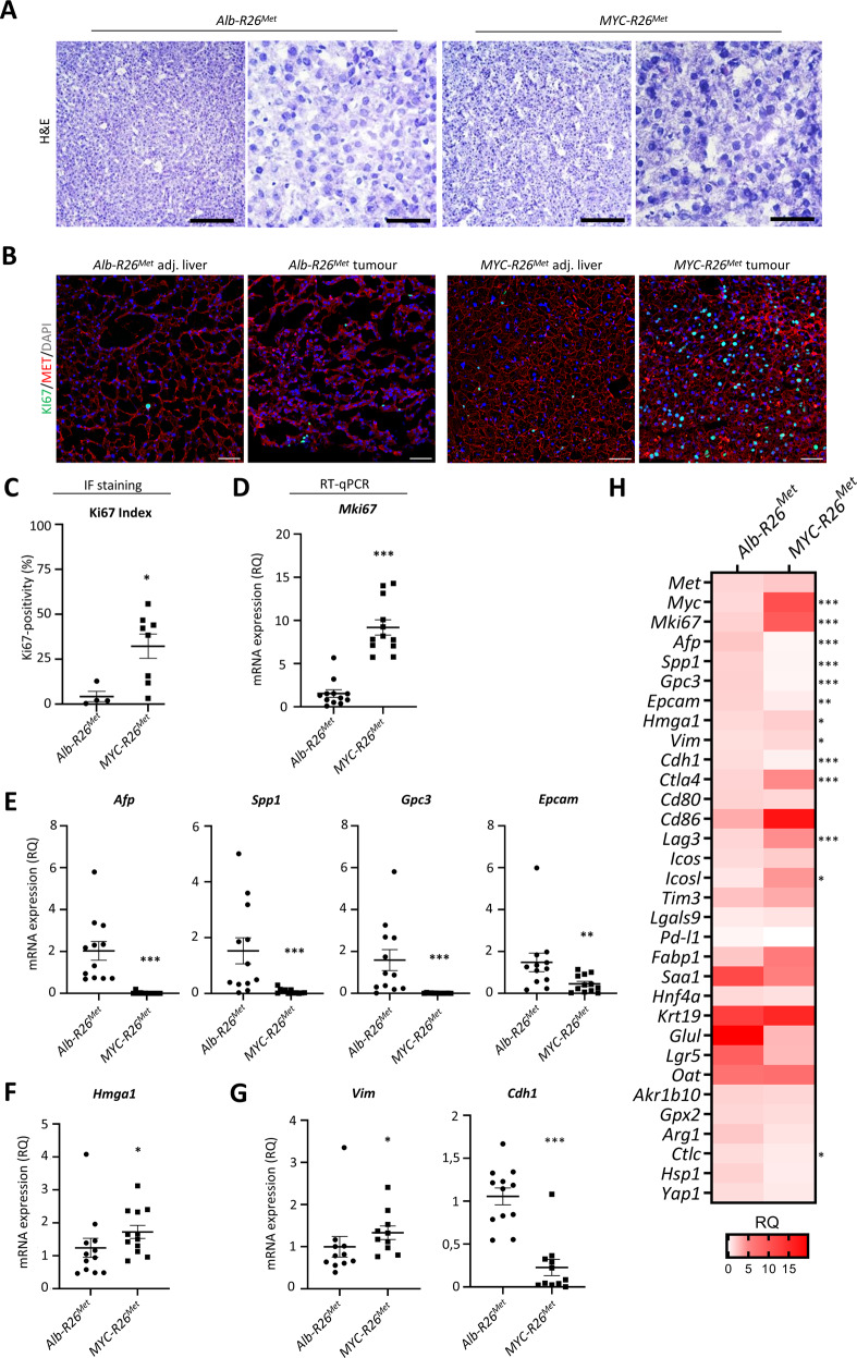
Fig. 3. Hepatocellular characteristics of Myc-R26Met and Alb-R26Met tumours.
A Representative haematoxylin and eosin staining of frozen, fixed Alb-R26Met and Myc-R26Met tumours and adjacent livers. B, C Representative images (B) and graph with quantifications (C) of Ki67 immunofluorescence staining of Alb-R26Met and Myc-R26Met tumour sections (scale bar: 50 µm). D–G mRNA expression levels by RT-qPCR of the proliferation marker Mki67 (D), of HCC markers Afp, Spp1, Gpc3, and Epcam (E), of the non-histone chromatin protein Hmga1 (F), and of mesenchymal Vim and epithelial Cdh1 markers (G) in Myc-R26Met versus Alb-R26Met tumours. H Heatmap reporting the RQ differential expression of all genes evaluated by RT-qPCR in this study. Values were normalised with the Gapdh housekeeping gene and expressed as RQ, all values relative to Alb-R26Met tumours. Statistical analyses were performed by Mann–Whitney. *p ≤ 0.05; **p ≤ 0.01; ***p ≤ 0.001.

