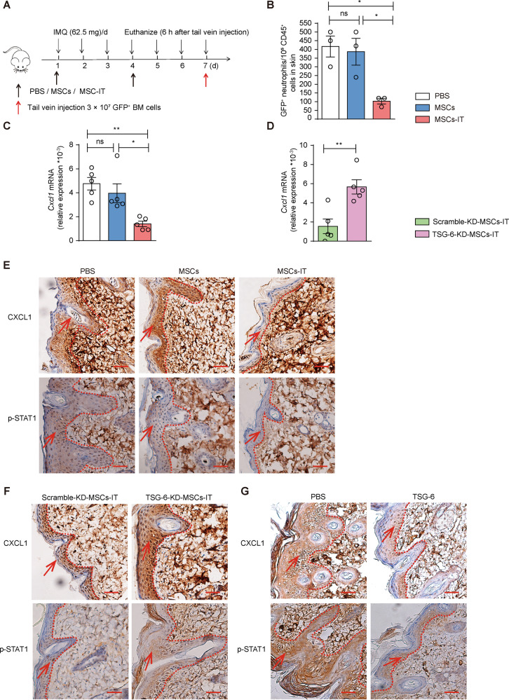
Fig. 6. MSCs-IT downregulated CXCL1 via TSG-6.
Bone marrow cells from GFP mice were infused into mice on day 7 of MSC treatment. Flow cytometry was used to analyze the proportion of GFP-positive neutrophils in the lesions 6 h later (n = 3 mice for each group). A The experimental scheme. B Enumeration of GFP positive neutrophils in the indicated groups, including IMQ + PBS group, IMQ + MSC group, and IMQ + MSC-IT group. In addition, we further examined the CXCL1 expression (n = 5 mice for each group). C The relative expression of Cxcl1 in the indicated groups. D The relative expression of Cxcl1 in the scramble-KD-MSC-IT group and TSG-6-KD-MSC-IT group. E–G The CXCL1 and p-STAT1 immunostaining of skin in the indicated groups. Scale bars: 50 µm. Data were shown as means ± SEM, *p < 0.05, **p < 0.01, ns: not significant.

