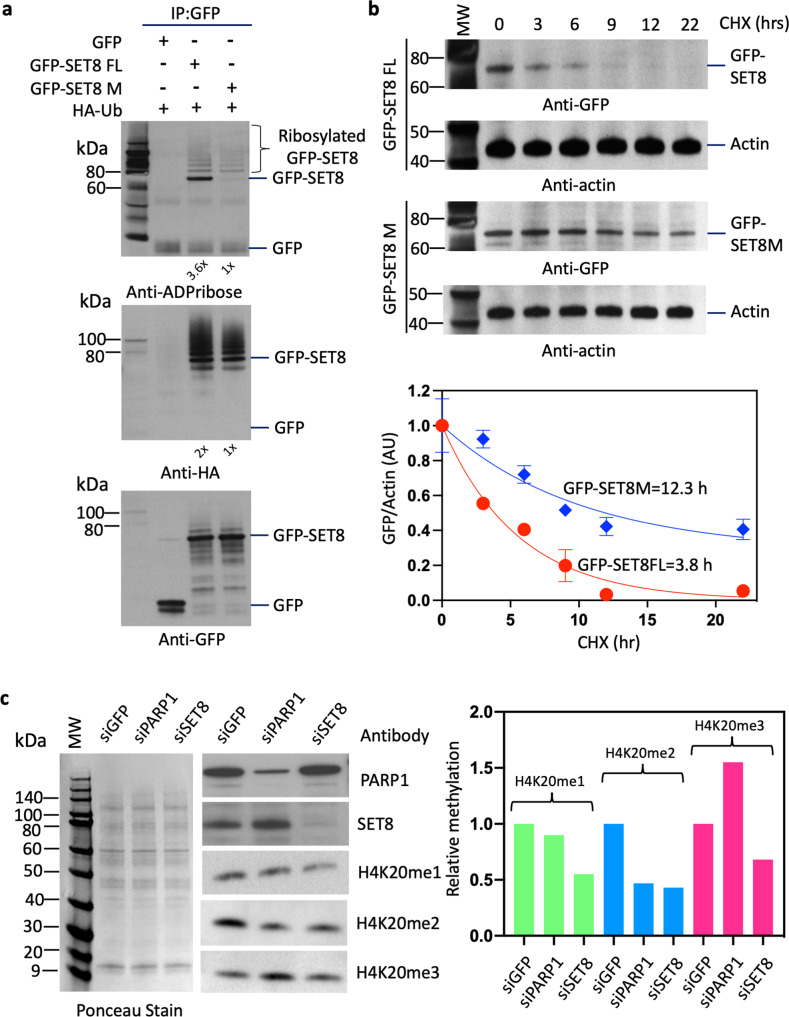
Fig. 4. PARP1 regulates SET8 protein stability.
a GFP-SET8 immunoprecipitation in overexpressed GFP-SET8 FL or GFP-SET8M with HA-Ubiquitin in COS-7 cells. Western blots detecting the amount of ADP-ribosylation (top panel), Ubiquitination (middle panel), and GFP fusion protein levels (bottom panel) in GFP immunoprecipitates. The fold increase was calculated by densitometry and indicated at the bottom of the western blot. b Cycloheximide chase analysis of SET8 stability in GFP-SET8 FL or GFP-SET8 M in HeLa cells. Western blots detecting the amount of GFP-SET8 FL and GFP-SET8 M protein levels with their respective actin levels (control) during cycloheximide time course (top panel). Respective densitometry analyses of GFP/Actin ratio representative of at least 2 biological experiments (bottom panel). c Western blots (left side) detecting the amount of PARP1 (top), SET8 protein (middle) as well as the amount of H4K20me1, H4K20me2, and H4K20me3 levels (bottom) in total protein extract of knockdown HeLa cells treated with siRNA (esiRNA) for GFP (control), esiRNA PARP1 and esiRNA SET8, respectively. Respective densitometry analyses of protein abundance representative of at least 2 biological experiments are shown (right side; n = 2). Ponceau stain was used as control (left side).

