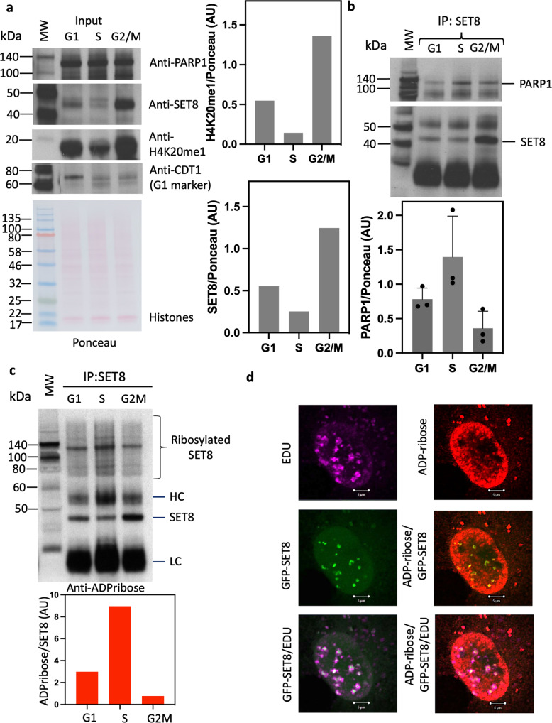
Fig. 5. Cell cycle-dependent interaction of PARP1 and SET8 correlates with global H4K20me1.
a Western blot indicating PARP1 (top, left), SET8 (middle, left), and H4K20me1 (middle, left) levels in total protein extracts from HeLa cells synchronized in G1, S, and G2/M phases, respectively. Western blot of CDT1 protein levels is shown as a cell cycle synchronization control as well as Ponceau stain for loading control and densitometry analyses (bottom, left). Respective densitometry analyses of H4K20me1 (top, right; n = 2) and SET8 (bottom, right; n = 2) relative protein abundances are shown (right) and representative of at least 2 biological experiments. b SET8 immunoprecipitation from total protein extract in HeLa cells synchronized in G1, S, and G2/M phases. Western blots detection of PARP1 (top, left) as well as SET8 immunoprecipitated protein levels (bottom, left) are revealed. Densitometry analyses of PARP1/SET8 ratio during G1, S, and G2/M cell cycle phases are shown (right; n = 3) and are representative of at least 2 biological experiments. c SET8 immunoprecipitation from total protein extract in HeLa cells synchronized in G1, S, and G2/M phases. Western blots detection of ADP-ribosylation as well as SET8 protein levels in SET8 immunoprecipitates (top panel). Respective densitometry analyses of SET8 ADP-ribosylation abundance representative of at least 2 biological experiments are shown (bottom panel; n = 2). d Pulsed chased cells with 5-ethynyl-2′-deoxyuridine (EdU) to label DNA (magenta) is transfected with GFP-SET8 (green). Endogenous ADP-ribose (red) is revealed by anti-ADP-ribose conjugated with Texas Red. Merged images demonstrate the colocalization of EDU, SET8 and ADP-ribose.

