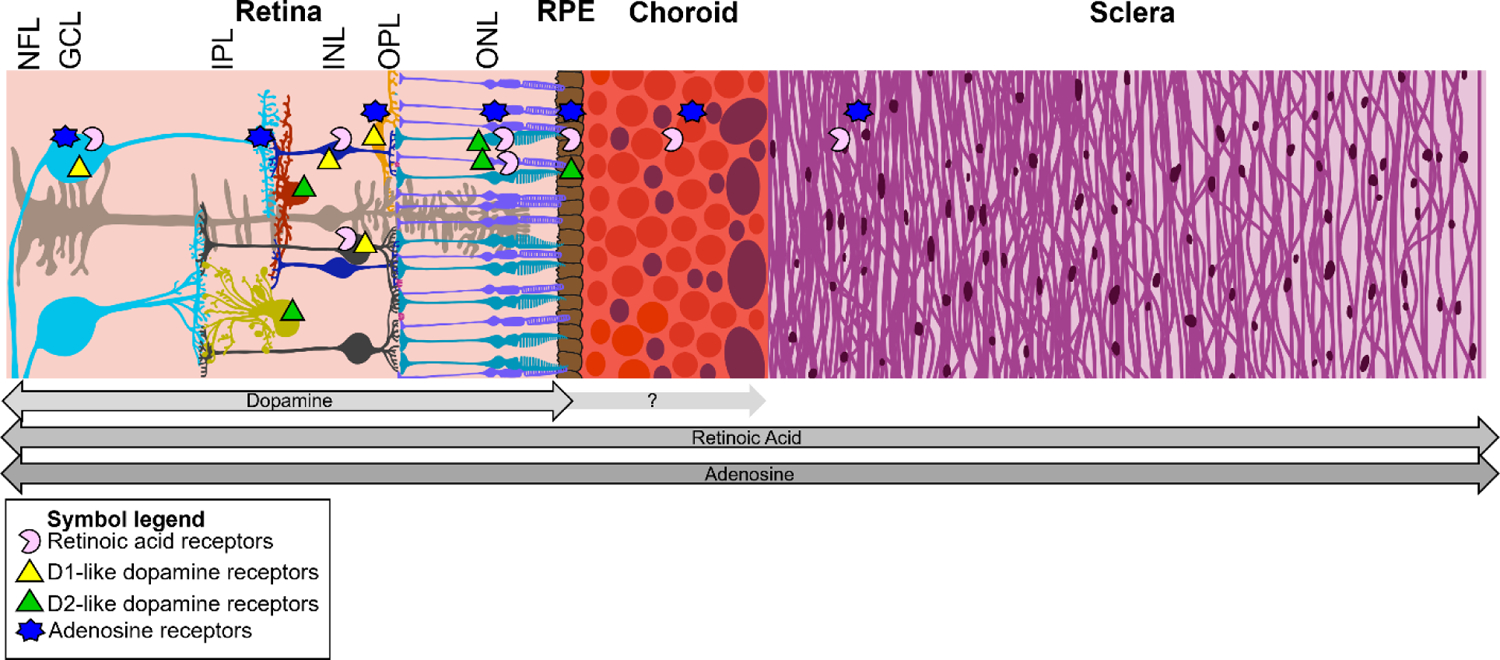
Figure 2:

Cartoon representation of the human posterior eye wall and the path of a hypothetical retinoscleral signal. Arrows below the diagram signify where each molecule has been implicated as a potential signaling molecule. The proportions of each layer are approximately to scale. NFL: nerve fiber layer, GCL: ganglion cell layer, IPL: inner plexiform layer, INL: inner nuclear layer, OPL: outer plexiform layer, ONL: outer nuclear layer, RPE: retinal pigment epithelium.
