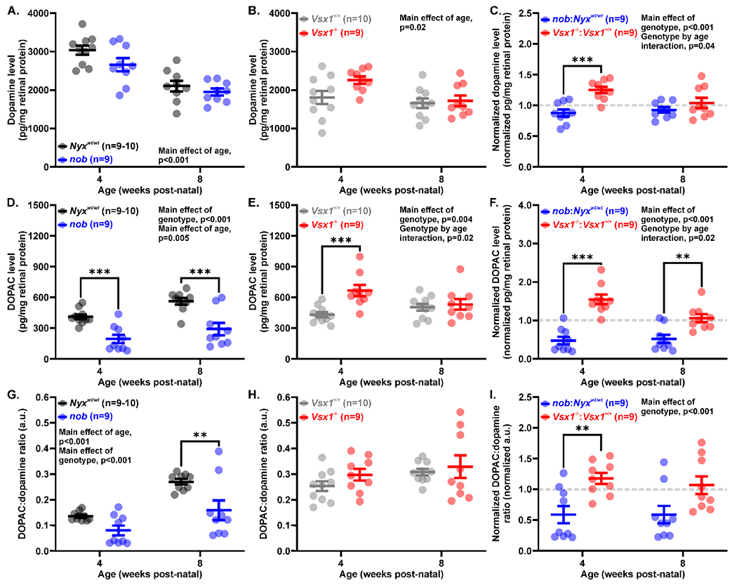
Figure 2.

Greater modulation in the retinal dopaminergic system in nob than Vsx1−/− mice. (A-B) Retinal dopamine levels measured with HPLC in (A) nob (blue) and Nyxwt/wt mice (black) as well as (B) Vsx1−/− (red) and Vsx1+/+ mice (gray). Retinal dopamine levels were unchanged in both genotypes, but both Nyxwt/wt and nob mice had reduced dopamine with age. (C) Dopamine levels normalized to wild-type average dopamine levels at each age. nob mice had significantly less retinal dopamine at 4 weeks post-natal than Vsx1−/− mice. (D-E) Retinal DOPAC levels measured in (D) nob and (E) Vsx1−/− mice as well as their wild-type counterparts. nob mice had reduced DOPAC levels at both ages, while Vsx1−/− mice had increased DOPAC at 4 but not 8 weeks post-natal. (F) Normalized DOPAC levels at 4 and 8 weeks post-natal. nob mice had reduced DOPAC levels compared to Vsx1−/− mice. (G-H) Ratio of DOPAC to dopamine levels, an indirect measurement of dopamine turnover, in (G) nob and (H) Vsx1−/− mice. nob mice had reduced dopamine turnover but Vsx1−/− mice did not. (I) Normalized DOPAC:dopamine ratio. Dopamine turnover was significantly more affected in nob than Vsx1−/− mice. Data are mean ± SEM and significance is *p<0.05, **p<0.01, and ***p<0.001. Comparisons were performed with two-way ANOVA (A-I) with Holm-Sidak multiple comparisons tests.
