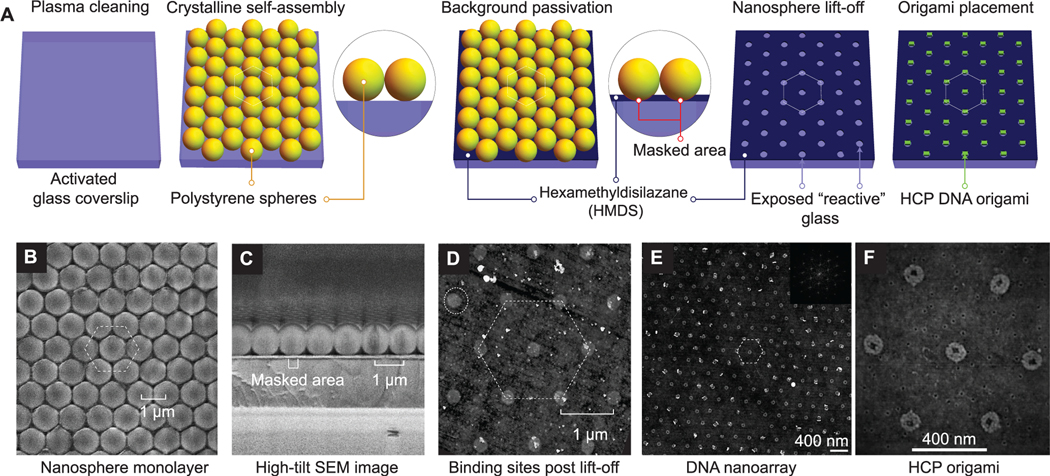
Figure 2.
Bench-top DNA origami nanoarray fabrication. (A) Schematic illustration of the DNA origami patterning process through 2D nanosphere close-packing, selective passivation, lift-off, and finally, Mg2+-mediated origami placement. (B, C) SEM images of nanosphere close-packing (top view and cross-section), respectively. (D) AFM images of binding sites. (E, F) AFM images of microscale origami placement. (inset) 2D FFT demonstrating close-packing. Experimental results demonstrate data analogous to schematic depiction (A) of process steps.

