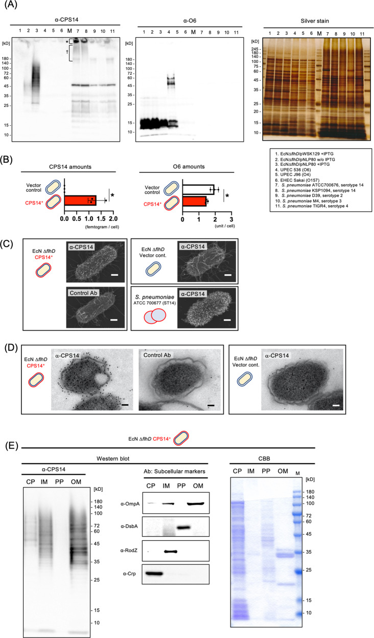
Fig. 2. Pneumococcal CPS expression on EcN ΔflhD cells.
A Western blot using anti-CPS14 (left panel) and anti-O6 antisera (middle panel), with sliver staining (right panel) as the loading control. The bacterial strains used in this experiment are shown with lane numbers at the lower right. For CPS14- and O6-probed western blot, the whole cells of all examined E. coli strains were standardized at an OD600 of 4.0, and the whole cells of all examined S. pneumoniae strains were standardized an OD600 of 8.0 using SDS-PAGE sample buffer. Twenty microliters of each sample was applied to the 12.5% polyacrylamide SDS-PAGE, electro-transferred onto a PVDF membrane, and probed with an anti-lipid A-core oligosaccharide antibody. For silver staining, 10-fold diluted whole-cell samples were prepared, i.e., whole cells of E. coli and S. pneumoniae were standardized an OD600 of 0.4 and 0.8, respectively, and twenty microliters of each sample was applied to the 12.5% polyacrylamide SDS-PAGE. Asterisk and dagger on CPS14-probed western blot membrane (left panel) denote CPS14-specific signals detected in the wells and the stacking gel of SDS-PAGE gel, respectively, shown in lanes 7 (S. pneumoniae, strain ATCC 700676 [serotype 14]) and 8 (S. pneumoniae, strain KSP1094 [serotype 14]). B Amounts of exogenous pneumococcal CPS14 (left panel) and E. coli original O6 (right panel) in whole cells of EcNΔflhD/pWSK129 (vector control, n = 3) and EcNΔflhD/pNLP80 (CPS14+, n = 3). All whole-cell samples used for analysis were collected from independent experiments. Data are expressed as the mean ± SD from results obtained in three independent experiments. *p ≤ 0.05, when performed with a Mann–Whitney U-test. C Immuno-FE-SEM. EcN ΔflhD cells harboring CPS14+ vector and the vector control, and S. pneumoniae strain ATCC 700676 (serotype 14) were probed with anti-CPS14 antibody (α-CPS14) or non-immunized rabbit serum (Control Ab). Primary antibodies were used at 1:500 dilutions. Anti-rabbit IgG antibody conjugated with 12-nm colloidal gold were used as the secondary antibody at 1:50 dilutions. Shown are representative images with scale bars indicated at the lower right. Bars: 200 nm. D Immuno-TEM. Whole cells of EcN ΔflhD harboring CPS14+ vector and the vector control were incubated with α-CPS14 or the control Ab. The primary antibodies were used at 1:500. Anti-rabbit IgG antibody conjugated with 12-nm colloidal gold was used as the secondary antibody at 1:50 dilutions. Shown are representative images with scale bars indicated at the lower right. Bars: 200 nm. E Subcellular fractionation of CPS14-expressing EcN ΔflhD cells. Subcellular fractions (cytoplasm [CP], inner membrane [IM], periplasm [PP], and outer membrane [OM]) were probed with α-CPS14, and with antisera against the following subcellular marker proteins: Crp [CP], RodZ [IM], DsbA [PP], and OmpA [OM]. Subcellular fractions were also stained with CBB. A 20-fold concentrated cytoplasmic fraction (CP), a 100-fold concentrated inner membrane fraction (IM), a 20-fold concentrated periplasmic fraction (PP), and a 100-fold concentrated outer membrane fraction (OM) were prepared from the bacterial suspensions standardized to OD600 = 2.0. Ten microliter of each sample was analyzed by SDS-PAGE.

