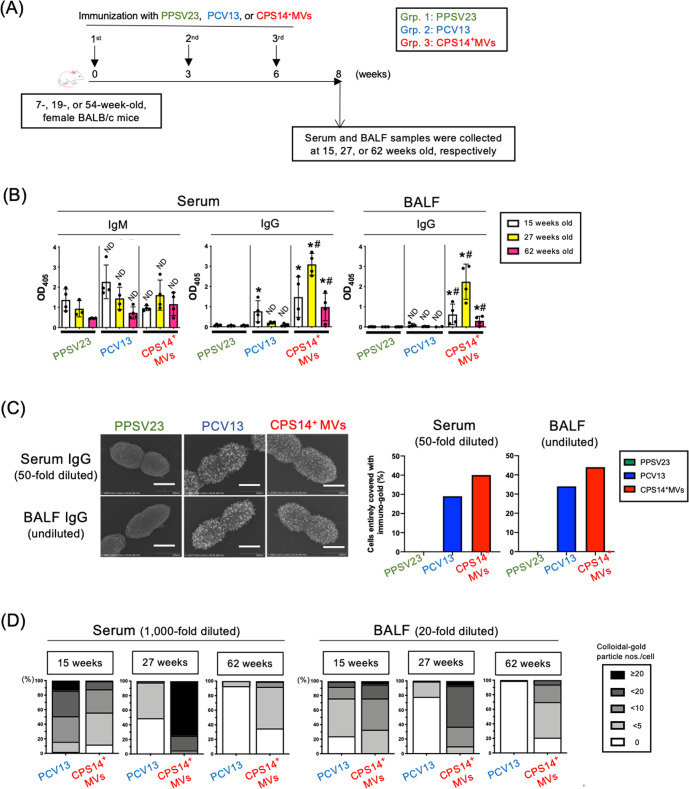
Fig. 6. Immunization study using mice at different ages.
A Timeline of immunization: CPS14+MVs vs. PPSV23 or PCV13 in mice at different ages. Thirty-six female BALB/c mice of different ages (7 weeks [n = 12], 19 weeks [n = 12], and 54 weeks [n = 12]) were subcutaneously immunized with PPSV23, PCV13, or CPS14+ MVs (PPSV23: n = 4 at each age, PCV13: n = 4 at each age, CPS14+ MVs: n = 4 at each age). B Humoral immune responses against CPS14. Serum IgM and IgG, and BALF IgG were examined. In ELISA for serum IgM and BALF IgG, the samples were used at 1:100 dilution. In ELISA for serum IgG, the samples were used at 1:1000 dilution. The results are expressed as OD405 values (mean ± SD) after a 30-min incubation with AP substrate. Shown are the results of statistical analysis using one-way ANOVA followed by Tukey’s multiple comparison test.ND: no statistically significant difference, when compared to Grp. 1 (PPSV23) of the same age. *p ≤ 0.05, when compared to Grp.1 (PPSV23) of the same age. #p ≤ 0.05, when compared to Grp. 2 (PCV13) of the same age. C Reactivity of mouse serum and BALF to pneumococcal cells. After analyzing a serotype-14 laboratory strain, ATCC 700676, by immuno-FE-SEM, reactivities of serum and BALF samples with the cells were evaluated. The samples prepared from all the mice in each group (n = 4) at the same volume were used for the immuno-FE-SEM. The 50-fold diluted serum mixture and the undiluted BALF sample mixture of 15-week-old mice were used. Shown are representative the electron-micrographs, which are merge of the secondary electron image and the reflection electron image. Scale bars indicated at the lower right. Bars: 500 nm. In addition, 100 cells were also randomly selected in each group, and then the cells that were fully covered with immuno-gold were counted. The results of serum (50-fold diluted) and BALF (undiluted) are indicated by two bar graphs in the right. D The samples prepared from all the mice in each group (n = 4) at the same volume were used for the immuno-FE-SEM using a clinical isolate of serotype 14 pneumococcus obtained from patients with. The 1:1,000 diluted serum mixture and the 1:20 diluted BALF sample mixture of 15-, 27-, and 62-week-old mice were used. In total, FE-SEM images of 100 cells were randomly captured for each group at 5 × 10⁵-fold magnification. A rectangular area defined as 180 × 250 nm2 was cropped from the center of a pneumococcal cell. The number of immuno-gold particles on the surface per cell were counted.

