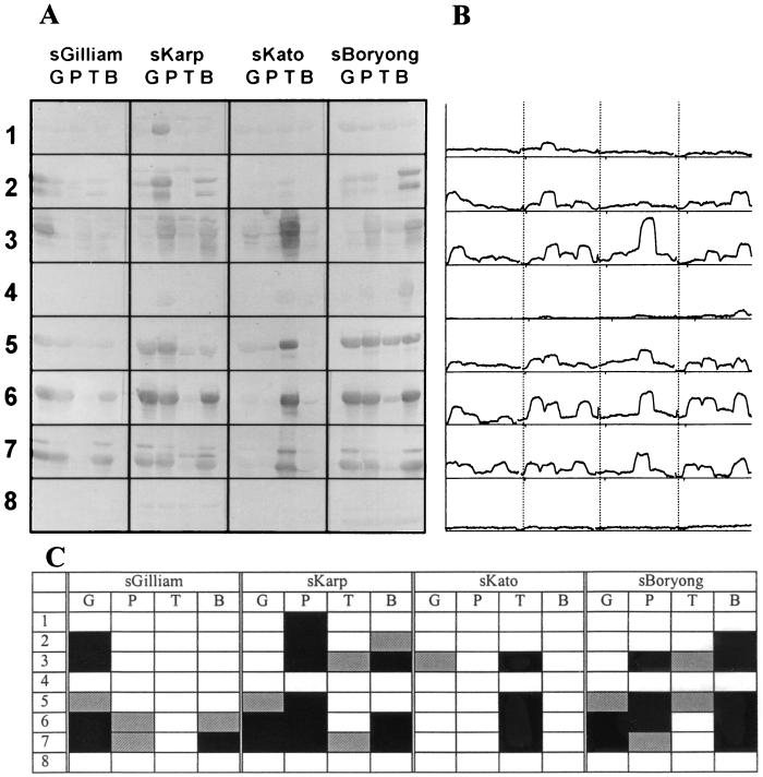
FIG. 2.
(A) Immunoblot of ΔTsa fusion proteins with sera from hyperimmunized mice. Induced fusion constructs were lysed, electrophoresed, transferred to nitrocellulose papers, and reacted with the indicated polyclonal sera (see below). Numbers indicate ΔTsa fragments shown in Fig. 1. (B) Densitometry analysis of the immunoblot. (C) Summary of immunoblotting analysis of sera from hyperimmunized mice with ΔTsa fragments. Black squares and gray squares indicate strongly positive and positive reactions (see the text), respectively. sGilliam, sKarp, sKato, and sBoryong, sera from mice immunized with Gilliam, Karp, Kato, and Boryong, respectively. G, P, T, and B, amino acid fragments derived from Gilliam, Karp, Kato, and Boryong, respectively.

