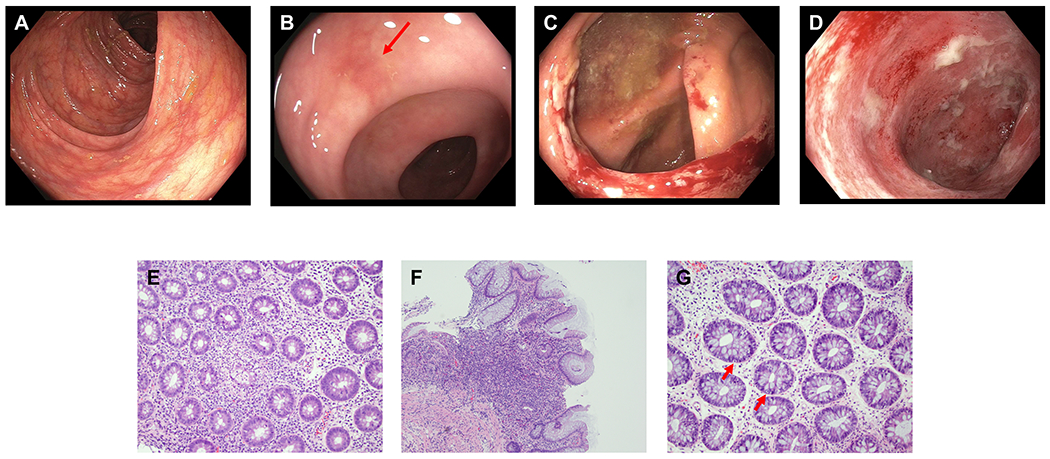
Figure 4.

Endoscopic and histologic findings of ICI colitis. A-D: Representative endoscopic images. (A) normal gross appearance; (B) focal erythema (red arrow); (C) focal ulceration; (D) diffuse inflammation. E-G: Representative histologic findings. (E) Active inflammation characterized by cryptitis and crypt abscess, magnification 100x; (F) Expansion of chronic inflammation in lamina propria, magnification 100x; (G) Intraepithelial lymphocytosis and increased crypt epithelial apoptosis (red arrows), magnification 200x.
