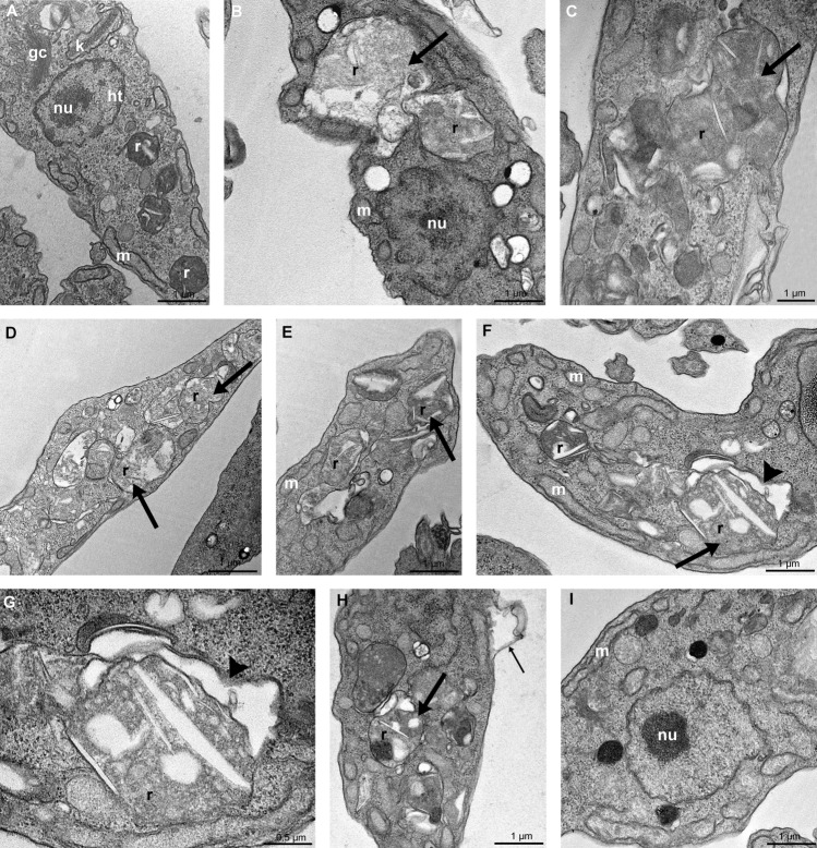
Figure 5.
Transmission electron microscopy of T. cruzi epimastigotes in the presence of FEX for 72 h. (A) Non-treated epimastigote with the Golgi complex (GC) near the bar-shaped kinetoplast (k), the nucleus with the condensed heterochromatin (ht) close to the nuclear envelope and around the nucleolus (nu), a single branched mitochondrion (m) and reservosomes (r) at the posterior end. (B–E) Treatment with 20 µM (B, C) and 30 µM (D, E) caused a loss of rounded shape and matrix content of the reservosomes (thick arrow). (F–I) Treatment with 40 µM also caused the detachment of the reservosomes from the cytoplasm (F, G, arrowhead), detachment of the plasma membrane (H, thin arrow), and the unpacking of nuclear heterochromatin (I).

