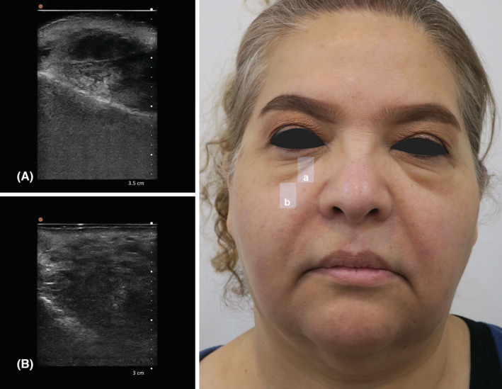
FIGURE 1.

(A) Ultrasonography of the right tear trough, B mode, sagittal view by handheld 20 MHz ultrasound; (B) Ultrasonography of the mid cheek in midpupillary line inferior to mid cheek groove, B mode, sagittal view, by handheld 20 MHz ultrasound. (A) It shows edema in periorbital area and hyper echogenicity in suborbicularis oculi fat (SOOF). (B) A mass about 20 mm in length has filled the whole space between the maxillary bone and skin and multiple bright hyperechoic spots with mini‐comet‐tail artifact in hypoechoic matrix are indicative of polycaprolactone deposits.
