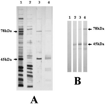
FIG. 1.
Characterization of the major surface antigens of M. agalactiae. (A) Total proteins (lane 1), soluble fraction (lane 2), and a Triton X-114 phase fraction (lane 3) were separated by SDS-PAGE, transferred to a nitrocellulose membrane, and India ink stained (11). Detergent-phase membrane-bound proteins (lane 3) were also immunostained with serum of a symptomatic naturally infected sheep (lane 4). (B) Immunoblotting of Triton X-114 phase fractionated proteins from M. agalactiae with preimmune sheep serum (lane 1) and after 9 days (lane 2), 24 days (lane 3), and 57 days (lane 4) of experimental infection (8).

